Article Text

Abstract
Background Translational research on clot composition may be advanced by the use of clot analogs for the preclinical evaluation of mechanical thrombectomy devices. This work describes a novel set of clot analogs to represent a diverse range of fibrin and red blood cell (RBC) compositions for use in acute ischemic stroke (AIS) occlusion models.
Method Fresh whole blood obtained from ovine species was used to create seven different clot analog types. Five replicates were formed for each clot type. Varying amounts of whole blood constituents were mixed with thrombotic factors to create clots of varying compositions. Following histological processing, five sections from each clot were stained with H&E and Martius Scarlet Blue. Fibrin, RBC and white blood cell compositions were quantified.
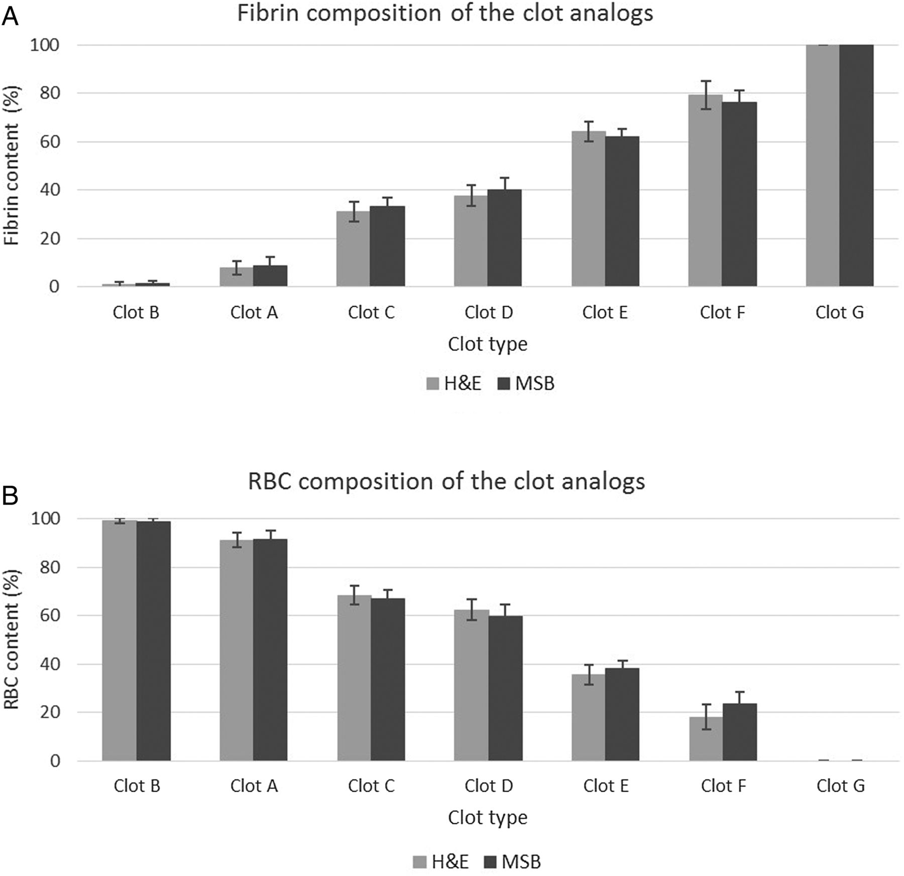
Results Histological examination demonstrated that the clot types had a distinct RBC and fibrin composition. No significant difference in composition was shown between replicates (p>0.05), indicating that the method of clot formation was reproducible. Percentage fibrin composition of the clot types was 1%, 8%, 31%, 38%, 64%, 79%, and 100%. A significant difference in fibrin and RBC composition between clot types was observed (p<0.05).
Conclusions Seven different clot types were developed to replicate common AIS thrombi. These clot analogs may be beneficial for the preclinical evaluation of endovascular therapies, and may be applied to interventional technique training.
- Thrombectomy
- Stroke
Statistics from Altmetric.com
Introduction
Recanalization and the restoration of downstream blood flow in the territory of a proximal cerebral artery has been established as the definitive treatment for acute ischemic stroke (AIS).1 Timely and effective recanalization of occlusive thrombi or clots in such arteries may be influenced by a number of underlying factors, including clot composition2 and type of occluded vessel.3 Mechanical thrombectomy (MT) has recently been established as an effective revascularization therapy for the treatment of AIS.4 ,5 Clinically relevant clot analogs are valuable for the pre-evaluation of MT devices, to assess the effect of various clot compositions on device performance. A common clot analog model is the Gralla model of thrombin induced clots prepared with radiopaque substances6 ,7 for clot visualization. This model is a red blood cell (RBC) rich clot,7 similar in composition to that formed by spontaneous coagulation.8 ,9 Another experimental approach is the preparation of a clot by sedimentation, with a multiple layered structure, consisting of a fibrin rich and RBC rich component.10 In order to simulate the physiological blood flow environment, Chandler11 proposed a closed loop technique to form a clot containing a large fibrin component in dynamic conditions.11–13
There are significant limitations associated with previously described clot models. Homogenous RBC dominant clots tend to be unstable and are susceptible to fragmentation during the MT evaluation procedure.2 Some clot models are prepared using radiopaque substances, such as barium sulfate,2 ,6 ,7 which significantly affects the clot's mechanical properties by decreasing elasticity.7 In addition, the histological characteristics are inconsistent with thrombi retrieved from AIS patients.14–18
A diverse composition of human AIS thrombi is documented in the literature,14–18 highlighting a clear need for clot analogs that cover a range of fibrin and RBC compositions. Based on a modification of existing11 ,13 ,19–21 and novel methodologies, this work describes the formation of seven clot analog types with varying fibrin and RBC compositions, intended to represent commonly retrieved AIS thrombi. The clot analogs may be used to expand our knowledge about the effect of thrombus composition on MT device performance, and may facilitate the development of new and refined techniques in the treatment of AIS. They may also be used for in vitro occlusion models for training in interventional techniques, and may facilitate the development of diagnostic imaging technologies to visualize clot composition in vivo.
Materials and methods
Blood collection
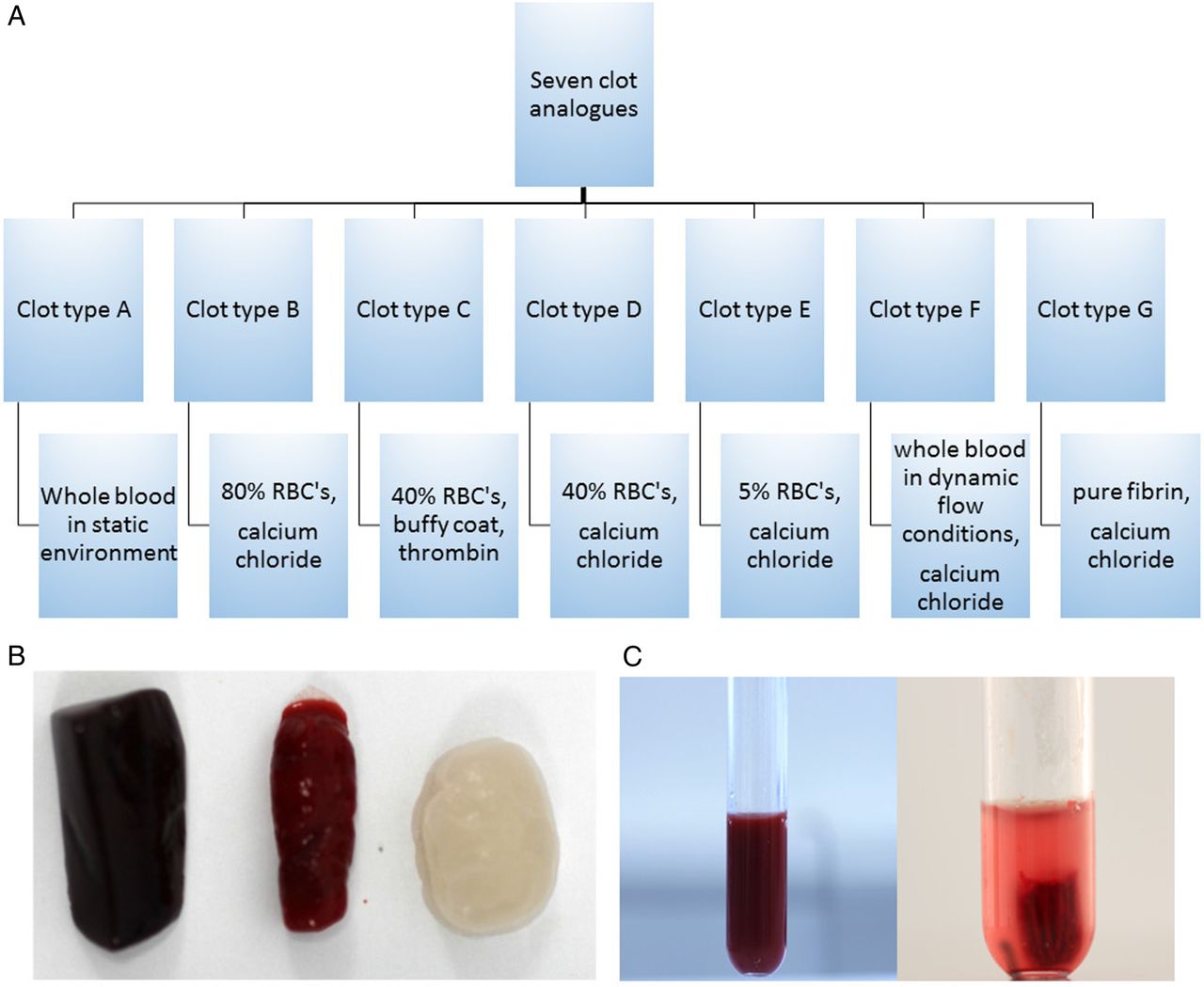
Blood was obtained from the jugular vein of ovine species (males and females; age range 1–7 years; weight range 70–120 kg) at a licensed facility (Ash Stream Ltd). Ovine blood was chosen because it has been demonstrated to be most suitable for coagulation studies,22 and venous blood was obtained to minimize stress to the animal. Seven different clot analog types were created, with five clot replicates formed for each type, as described below and illustrated in figure 1A. For clot types B–G, blood was anticoagulated in 3.2% sodium citrate solution in a 9:1 ratio immediately after collection (mixing ratios were measured per volume). To separate the whole blood constituents, centrifugation was performed at 550 g for 15 min. Coagulation was generally initiated following addition of 2.06% calcium chloride solution to the blood components in a 1:9 ratio, except where otherwise stated. Clotted material was allowed to mature for approximately 30 min to 1 h at 37°C.

(A) Formation of each clot type formed generally by mixing different thrombotic factors (calcium chloride unless otherwise stated) with varying blood constituents. (B) Gross images of clots: spontaneously formed clot type A (left), clot type D (middle), and fibrin rich clot type G (right). (C) Coagulated clot type D (left) and retracted clot type C (right). RBCs, red blood cells.
Clot formation
Clot type A was formed in a static environment. Spontaneous coagulation was initiated by collecting whole blood into a syringe, left stationary for approximately 12 h at room temperature. Clot type B, a RBC rich clot, was formed by mixing citrated plasma and RBCs in a 1:4 ratio (ie, 80% RBCs) and subsequently coagulated. Clot type C was prepared using thrombin, and platelets were included to promote clot retraction.20 Prostaglandin E1 solution (final concentration of 500 ng/mL based on the study by Katori et al20) was added to citrated blood prior to centrifugation to minimize platelet activation during the centrifugation process. The extracted buffy coat and plasma were mixed in a 1:2 ratio, and subsequently mixed with RBCs in a 6:4 ratio (ie, 40% RBCs). Adenosine diphosphate 1 mM was added to reverse the effects of prostaglandin E1.20 Adenosine diphosphate was mixed with thrombin (final concentration 35 IU/mL blood) in a ratio of 0.16:1, before mixing with blood in a 1:7.75 ratio. The mixing ratios are based on the authors' experimentation. The high concentration of thrombin used is based on a study that demonstrates the formation of thin fibrin strands with high thrombin concentrations.19 Clot types D and E were formed by mixing citrated plasma with RBCs in a ratio of 6:4 (ie, 40% RBCs) and 19:1 (ie, 5% RBCs), respectively, and subsequently coagulated. Clot type F was prepared using a modification of the Chandler loop technique.11 ,13 ,21 Silicone tubing with an inner diameter of 7 mm and approximately 30 cm in length was mounted in a multi rotator device (Grant Bio PTR-35), and partially filled with approximately 10 mL of citrated whole blood. Calcium chloride was injected into the tubing, and the ends of the tubing were fixed to form a closed loop. The tubing rotated at a rate of 25 rpm for approximately 1 h (the speed was calculated based on the carotid blood flow rate of approximately 240 mL/min23). Clot type G, a fibrin rich clot, was prepared by coagulating citrated plasma in the absence of RBCs and buffy coat.
The RBC compositions of clot types B, C, D, and E were chosen based on the RBC composition of retrieved human thrombi reported in the literature.14–18 These data show that each retrieved thrombus has a unique composition, and there is great variation in RBC and fibrin content. It was not possible to replicate the composition of every human thrombus without having an excess number of clot analogs. Therefore, in this study, a number of selected concentrations were chosen that covered a wide range of RBC compositions.
Histological assessment
Immediately after clot formation, each of the clots were fixed in 10% neutral buffered formalin for 24–48 h. Following histological processing, they were embedded in paraffin wax and sectioned at 4–5 µm. Ten sections from each of the five clot analog replicates were obtained. Subsequently, five sections were stained with H&E (n=25 sections/clot type) and five with Martius Scarlet Blue (MSB) (n=25 sections/clot type). Histological evaluation was performed at the Galway-Mayo Institute of Technology (SD) and Beaumont Hospital by an experienced neuropathologist (MF). Sections were photographed using an Olympus BX41 microscope with an Olympus digital camera (model DP25) attached. Image J software (V.1.49v, National Institutes of Health, USA) was used for quantitative analysis of the percentage fibrin, RBCs and white blood cells (WBC's) by area. There is no standard technique available to analyze and quantify thrombi. The same quantification methodology was applied to assess the composition of the proposed analogs as reported previously to quantify the composition of human thrombi.14 Clot G was not subjected to image J analysis because fibrin was the only component visible microscopically.
Statistical analysis
The percentage composition of fibrin, RBCs, and WBCs were recorded in Microsoft Excel 2013, and statistical analysis was performed using Minitab 17 (V.17.2.1, Minitab Inc statistical software package). Summary data are expressed as percentage area.
To test the reproducibility of the applied technique, percentage composition of the clot analog replicates belonging to a specific clot type were compared using a one-way ANOVA. The relationship between different clot types was investigated using a two sample Student's t test. H&E and MSB staining were compared using a paired t test. For all statistical tests, statistical significance was noted at p<0.05.
Results
The fibrin and cellular composition of each of the seven clot types were quantitatively evaluated using histological techniques. Five sections from each clot were stained as described above to evaluate their histological composition. Figure 1B illustrates the gross appearance of three different clot types. Clot type C underwent significant retraction resulting in a marked reduction in clot volume (figure 1C).
Figure 2 illustrates the histological staining of different clot types. Clot A formed after spontaneous coagulation of whole blood (figure 2A) was replete with RBCs. Thrombin induced clot type C also revealed a predominance of RBCs with randomly distributed fibrin strands (figure 2B). Clot F formed in dynamic flow conditions had a predominance of fibrin and defined areas of RBCs and WBCs (figure 2C). The fibrin rich clot type G demonstrated a fine delicate fibrin network, as shown in figure 2D.

H&E (left) and Martius Scarlet Blue (MSB) (right) staining of four different clot types (×20 magnification). MSB stained fibrin red and erythrocytes yellow: (A) clot A replete with red blood cells (RBCs). (B) Thrombin induced clot C predominantly composed of RBCs with interspersed bands of fibrin. (C) Dynamically formed clot F, demonstrating a predominance of fibrin with collections of RBCs. (D) Fibrin rich clot, clot G, demonstrating delicate fine fibrin strands (magnification bars=100 µm).
Table 1 summarizes the fibrin and cellular compositions for all clot analog types based on the area analysis of the histologically stained slides. There was no statistical difference (p>0.05) in composition among the five clot replicates belonging to a specific clot type. This demonstrates that the technique employed for the clot formation is reproducible. Clot G had the highest fibrin content (100%±0%) with no RBCs present, while clot B had the lowest fibrin composition (0.95%±1.14%). In clots where RBCs were present, there was a predominance of RBCs observed in clot B (99.05%±1.14%) while clot F contained the lowest percentage of RBCs (18.07%±5.28). The fibrin and RBC compositions quantified by both staining techniques were comparable, as illustrated in figure 3.
Fibrin and cellular composition of the seven clot types stained with H&E and Martius Scarlet Blue, based on the area analysis of the stained sections
(A) Percentage fibrin composition of the seven clot types. No significant difference was observed between the two stains (p>0.05) for each clot analog type, based on a paired t test. (B) Percentage red blood cell (RBC) composition of the seven different clot types. No significant difference was observed between the two stains (p>0.05) for each clot analog type based on a paired t test. MSB, Martius Scarlet Blue.
Table 2 shows the percentage area variation of the fibrin, RBC, and WBC compositions between different clot types based on a two sample Student's t test at the 95% CI. There was a significant difference in fibrin and RBC percentage composition between clot types (p<0.05).
Percentage area variation of fibrin, and red blood cell and white blood cell composition between different clot types based on a two sample Student's t test at 95% CI
Discussion
This study successfully created seven distinct clot analog types in vitro that provides a platform for subsequent translational research on stroke clot composition. Studies on human thrombi retrieved from AIS patients have revealed a varying composition and morphology.14–18 While it is not possible to replicate the morphologic characteristics of every retrieved thrombus, a set of clot types have been proposed with a range of compositions (table 1). The clot analogs in this study range from very high RBC content to fibrin rich clots, and are clinically relevant as they are compositionally and histologically similar to thrombi retrieved from AIS patients.14–17 They are also reproducible and relatively simple to form.
Clot A formed in a static environment was replete with RBCs with a low fibrin composition and is in keeping with previously published studies.8 ,9 ,24 Varying the amounts of RBCs (clot types B–E and G) had a predictable effect on the amount of visible fibrin in the clot. Clot type F was formed with a predominance of fibrin, with clumps of RBCs and a cellular component (figure 2C). This clot type was formed using the Chandler loop technique,11 where a chamber of air remains on top of a vertical rotating circular loop (ie, the fluid is stationary and the tube is rotating). In contrast with clot type A formed under static conditions, the flow conditions for clot type F mimics the carotid blood flow rates in vivo (approximately 240 mL/min23). The fibrin rich pattern in clot type F is in keeping with the composition of clots formed in a dynamic flow model where clots formed in flow are firmer and more elastic,13 in comparison with RBC dominant clots formed using the Gralla method.6
Clot C was a retracted clot with a slightly lower fibrin composition in comparison with clot D, formed using the same ratio of plasma/RBCs. The small difference in fibrin composition may be explained by the presence of thrombin in clot C. High concentrations of thrombin promote the formation of thinner fibrin strands,19 and form a dense clot with a tighter fibrin network.25 Niessen et al26 demonstrated that thrombin induced clots are elastic and are associated with greater recanalization times during MT evaluation which may be possibly explained by the mechanical characteristics of the clots. Mechanical studies are required to delineate differences in the mechanical properties between clot C and clot D.
Thrombus morphology research on human AIS patients has revealed a variety of different thrombi compositions such that no two thrombi are histological identical, with fibrin and RBC composition spanning a comprehensive range.14–18 Retrieved human thrombi varied between fibrin rich, RBC dominant, and mixed thrombi. This research highlights the requirement for an experimental clot analog model with a great variety of fibrin and RBC compositions, and to evaluate MT devices under challenging and reproducible conditions. At present, however, the majority of clot models used for MT evaluation are RBC dominant clots.27 ,28 The performance of MT devices is influenced by the composition of the occluding thrombus. Vessels occluded with fibrin rich thrombi require a greater number of attempts at MT to achieve recanalization in comparison with RBC rich thrombi, and are consequently associated with a greater mean recanalization time.2 Animal models have also shown a lesser treatment effect of fibrin rich thrombi to tissue plasminogen activator, whereas RBC rich clots showed higher response rates.26 ,29 In fibrin rich clots, the nature of fibrin and the greater volume of fibrin requiring lysis may reduce the permeability of tissue plasminogen activator. We hypothesis that clots with high RBC content have a sparser underlying fibrin matrix, and they are more prone to fragmentation.2 The performance of MT with various clot analogs is beyond the scope of this study, but these clot analogs will form the basis for future studies on thrombolytics and MT efficacy, and determine the optimum thrombectomy technique to apply with a specific clot composition. The clots proposed have distinctly different compositions, resulting in different physical properties. We have observed that clots with high RBC content (eg, clot types A, B, C, and D) may be prone to fragmentation, and an effective MT technique should mitigate distal or new territory embolization. Because fibrin rich clots are generally resistant to thrombolysis, clot types E, F, and G may be more suited to MT.
An advantage of the proposed clots is that they do not contain substances which do not naturally form in vivo, and therefore lend themselves to use in imaging studies (CT or MRI) with less potential for artifacts. A combination of future imaging and more in depth knowledge about the effect of different clot compositions on endovascular therapies could become helpful to assign patients to a specific treatment strategy.
The authors acknowledge that some limitations to this study exist that merit consideration, in particular the exclusion of WBCs in some clot types, and the presence of platelets was not investigated. Correlation studies linking RBC/fibrin composition and stroke etiology have been documented16–18 but it is not clearly understood if WBC's and platelets are associated with stroke etiology. A further limitation of this study is the absence of calcified material in the clot analogs. However, only a small percentage of human AIS thrombi are calcified,7 ,30 suggesting it is not a common pathogenic process for AIS. It is unknown if the clot composition is altered over time because they were not tested histologically at different time points. From the authors' experience, the clots can be used for up to 3 weeks after formation when stored at 4°C. Finally, the mechanical characteristics of these clots have not been investigated, and this is the focus of future studies, in addition to assessing platelet composition using immunohistochemical techniques. Clot analogs for use in AIS models should ideally resemble the histological as well as the mechanical properties of human AIS thrombi.
Although the clot types described in this study represent some commonly retrieved human thrombi, composition of thrombi from patients with cardiovascular risk factors (diabetes, smokers, hypertension, and obesity) are not specifically represented in this study and other published studies describing clot analogs. There is no large study that focuses on the composition of thrombi from this cohort. If these data were available, the methodology used in this study could be applied to produce clinically relevant analogs focusing on the specific cohorts of AIS patients. Patients with cardiovascular risk factors are at an increased risk for developing atherosclerosis due to damage to the blood vessels/endothelial dysfunction. Atherosclerotic plaques have previously been described as having a lipid core and calcifications,31 ,32 which makes these clot types more complex in nature and clot analog formation more difficult.
Conclusion
This study outlines the formation and characterization of a range of clot types with varying fibrin and RBC compositions, intended to represent the diversity of histological patterns of AIS thrombi. The clot types may have value for practice and future research, in particular with the design of more clinically relevant models for the preclinical evaluation and development of MT devices, and for interventional technique training with such devices.
Acknowledgments
The authors acknowledge Mahmood Mirza for his assistance with editing the manuscript.
References
Footnotes
Contributors SD was involved in the study design, histological and statistical analysis, data interpretation, and manuscript writing. MF was involved in the interpretation of the results, and provided guidance and direction on drafting the manuscript. KM contributed to the design of the study. JT and DV were involved in the initial concept of forming clot analogs. DSL contributed to the design of the study, and provided guidance and direction on drafting the manuscript. ER and EM were involved in the design of the study. LM assisted with the statistical analysis. MG contributed to the initial concept of forming clot analogs, and was involved in the study design. All authors provided suggestions and feedback on the manuscript, and approved the final manuscript.
Funding This work was supported by Neuravi Ltd and the Irish Research Council grant No EBPPG/2014/50.
Competing interests SD reports grants from the Irish Research Council, during the conduct of the study, and reports financial relationships with Neuravi Ltd, outside the submitted work. KM, DV and MG report financial relationships with Neuravi Ltd, outside the submitted work. JT is a member of the scientific board for Neuravi Ltd. DSL reports grants from NIH-NINDS, Stryker, and Medtronic, outside the submitted work.
Provenance and peer review Not commissioned; externally peer reviewed.
Data sharing statement The authors agree to share any unpublished data on a request basis to the corresponding author.