Article Text

Statistics from Altmetric.com
Since the start of the COVID-19 pandemic, there has been an urgent need to develop protective measures to ensure patient and healthcare worker safety in clinical situations.
Direct/monocular ophthalmoscopy is particularly difficult to perform safely, given the very close proximity of the patient and the clinician. However, ophthalmoscopy is an important and on occasion an irreplaceable element of the neurological examination. It is particularly important for identifying papilloedema in headache, optic disc oedema and/or other retinal findings in acute visual loss, temporal disc atrophy in suspected multiple sclerosis or simply for reassuring someone with primary headache where the examination is otherwise normal or there are only superficial optic nerve drusen.1
Using an ophthalmoscope when wearing adequate personal protective equipment can be challenging. From our experience, it is impractical to visualise the optic fundi while wearing protective glasses or a face shield, and such attempts can mislead, contributing to diagnostic errors/or unnecessary investigation.
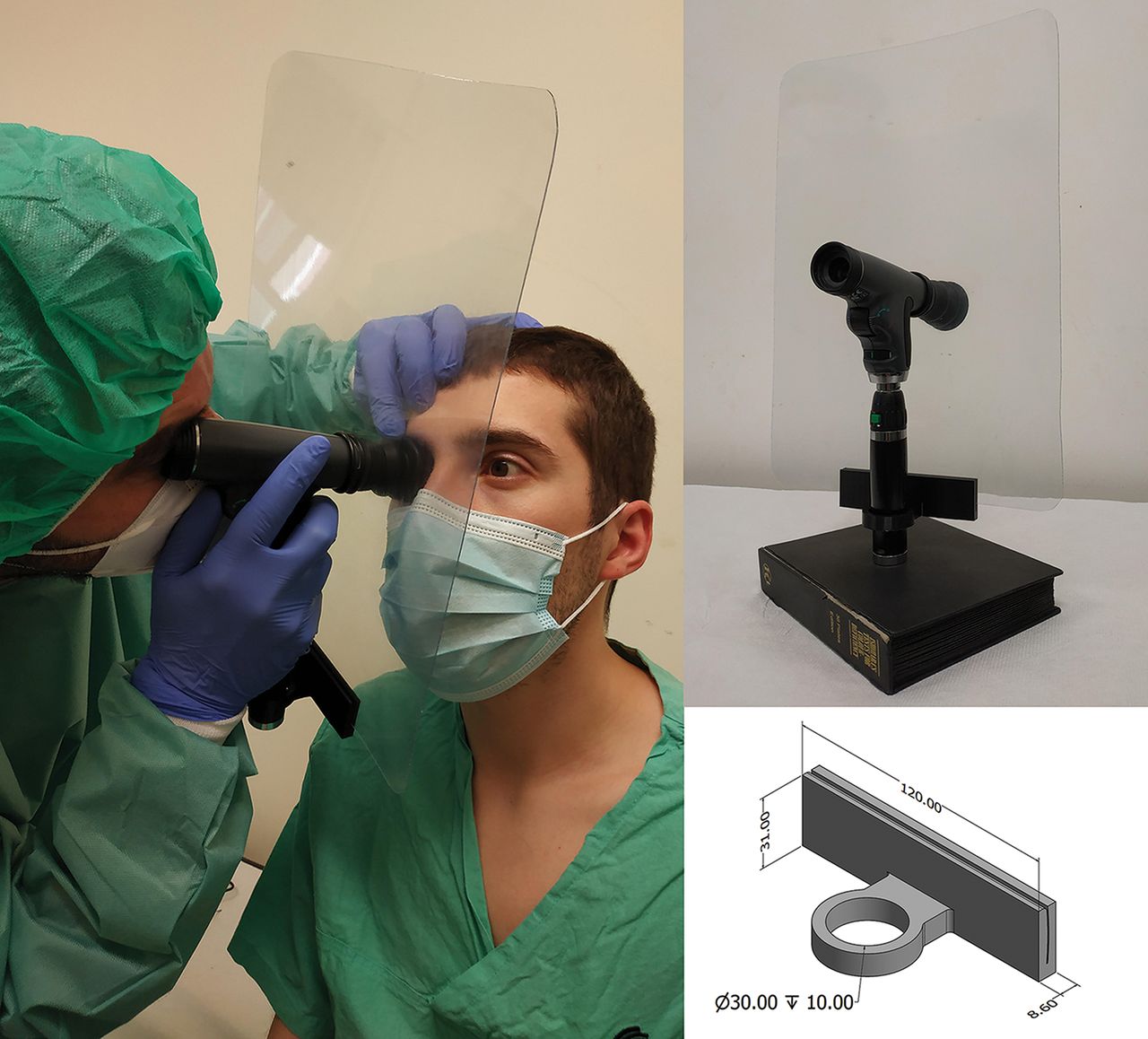
We, therefore, aimed to develop a protective shield that could be adapted to the ophthalmoscope and provide a safer but still effective ocular fundus observation. In collaboration with the Physics Department of University of Coimbra, and in accordance to recent publications on other protective materials in the ophthalmology,2 we developed a protective shield adapted to the Welch Allyn PanOptic ophthalmoscope (figure 1). The shield is made from common resistant and washable materials, allowing for home-made production (see figure 1 legend for specifics of materials and dimensions). The shield and the ophthalmoscope are cleaned using 70% alcohol wipes, allowed to air dry between each patient and washed with water detergent solution at the end of each shift.

Ophthalmoscope protective shield. The protective shield (left and right upper segments) comprises three elements: (1) a plexiglass sheet (37×26 mm, 0.6 mm thickness) with a central hole (32 mm diameter), (2) two rubber rings (O-rings, 31 mm inner diameter) encircling the ophthalmoscope’s head on each side of the sheet hole, (3) a three-dimensional printed polylactic acid component (right lower segment) docked around the power source/grip and attached to the lower border of the sheet to increase stability. The dimensions presented in computer-aided design model are in millimetres (right lower segment). We are currently making developments to the polylactic acid component to accommodate power handles of different sizes and simultaneous use of a charger.
Our routine sequence inside the room is ophthalmoscopy, followed by gloves doffing, hand hygiene, goggles donning and gloves donning, followed by the remaining examination. Importantly, the shield does not interfere with the technique or compromise diagnostic accuracy. At this point, we can recommend its use only for COVID-19 low-risk patients/environment and not for patients with COVID-19 infection, since its use precludes the simultaneous use of face shield or goggles. Note that the shield is for use with the PanOptic ophthalmoscope and cannot easily be adjusted for a standard hand-held ophthalmoscope.
This device has not been formally tested but applies the principles used in other personal protective equipment and is in line with current advice. We hope that sharing this information will prove helpful.
Footnotes
Correction notice This article has been corrected since it was published. Affiliation 4 has been amended to the correct site of the laboratory. 'New University of Lisbon' has been changed to 'University of Coimbra'.
Contributors AJ contributed to acquisition, analysis and interpretation of the data and drafting of the manuscript. AIM contributed to acquisition, analysis and interpretation of the data. MP, CN, JC and PGV contributed to concept and design, and critical revision of manuscript for intellectual content. JL contributed to acquisition, analysis, and interpretation of the data, drafting of the manuscript, study supervision, concept and design, and critical revision of manuscript for intellectual content.
Funding The authors have not declared a specific grant for this research from any funding agency in the public, commercial or not-for-profit sectors.
Competing interests None declared.
Patient consent for publication Not required.
Provenance and peer review Not commissioned. Externally peer reviewed by Nick Davies, London, UK.
Other content recommended for you
- Drusen and the misleading optic disc
- Use of personal protective equipment against coronavirus disease 2019 by healthcare professionals in Wuhan, China: cross sectional study
- Donning and doffing of personal protective equipment protocol and key points of nursing care for patients with COVID-19 in ICU
- Visual field constriction in a 42 year old woman
- OCT angiography in optic disc drusen: comparison with structural and functional parameters
- How to rapidly design and operationalise PPE donning and doffing areas for a COVID-19 care facility: quality improvement initiative
- Enhanced depth imaging optical coherence tomography of the optic nerve head improves correct diagnosis in glaucoma suspects without glaucomatous optic disc morphology
- Retinal imaging: what the neurologist needs to know
- Distinguishing optic disc drusen from papilloedema
- Inner retinal degeneration associated with optic nerve head drusen in pseudoxanthoma elasticum