Article Text

Statistics from Altmetric.com
Tick, tick… boom?
Often the people with most ‘need’—comorbidities and complications—cannot access all the diagnostic help available. Such is the problem with the concerns regarding ‘non-MRI conditional’ implanted cardiovascular devices and MRI. A Fo Ben reads with interest, therefore, about an American registry of 1.5-Tesla MRIs where 1000 people had a pacemaker and 500 had an implantable cardioverter–defibrillator (ICD). In total, there were 1499 lethal explosions… no, no sorry—I mean nothing happened. Well, nothing really. In one case (where the pre-MRI programming protocol was ignored), the ICD generator could not be interrogated afterwards, and there were six cases of self-terminating atrial fibrillation or flutter and six cases of partial electrical reset. Demonstrating that there was no device or lead failure in any patient with a non-MRI conditional pacemaker or ICD undergoing a nonthoracic MRI at 1.5 Tesla (who had the device programmed per the protocol) is a great boon clinically.
N Engl J Med 2017;376(8):755-764.
Memory almost full
The spirit of co-operation continues with this genome-wide association study of hippocampal volume from Nature Communications. Environmental factors, such as stress, affect the hippocampus, but genetic differences across individuals account for most of the population variation in its size and hippocampal size is a highly heritable trait. Hippocampal structural abnormalities are seen in mood disorders (such as bipolar), developmental disorders (such as schizophrenia), degenerative disorders (such as Alzheimer’s) and disorders with an unclear aetiology. A total of 33 356 individuals were studied, yielding six independent loci—three of which were in genes (ASTN2, DPP4 and MAST4). They also identified that genetic variants linked to smaller hippocampal volumes are associated with an increased risk of Alzheimer’s disease.
Nat Commun 2017;8:13624.
A strong cup of T?
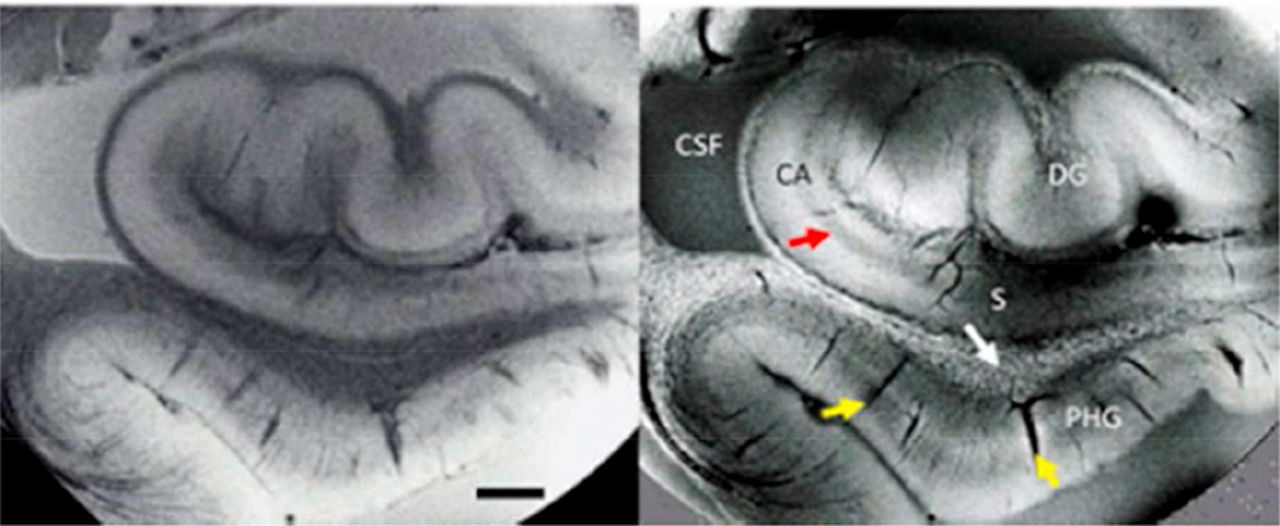
Anyone feeling emboldened about the safety of devices in a 1.5-Tesla MRI (above) may want to consider the power of superconducting materials that permit ever greater improvements in magnetic flux density. Budinger and Bird describe the potential for MRI and MR spectroscopy at 14–20 Tesla fields. Although the physiological and safety of these field strengths are discussed at length, A Fo Ben notes that the exquisite images (figure 1) are taken from ex vivo tissue—neatly side-stepping some of the safety fears regarding acoustics, projectiles and overheating.
Neuroimage 2017 pii: S1053-8119(17)30 090–3.
Susceptibility-weighted MRI of ex vivo hippocampal tissue. Signal amplitude (left) and phase (right) show distinctly different contrast. The dentate gyrus (DG), cornu ammonis (CA), subiculum (S) and parahippocampal gyrus (PHG) are readily identifiable. Contrast noted around vessels (yellow arrows), pyramidal cell layers (red arrow) and in white matter (speckles). CSF, cerebrospinal fluid.
Sighs matters
A sigh is just a sigh? How much do you really know about the control circuit for sighing? Small and specialised subpopulations of nerve cells in a breathing centre (retrotrapezoid nucleus/parafacial respiratory group) express neuropeptide genes for neuromedin B or gastrin-releasing peptide. These cells project to the preBötzinger complex, the respiratory rhythm, generator, which has complementary receptors for the peptides. The preBötzinger complex is key to inspiration, and a ‘double burst’ from this area induces the prolonged inspiration needed to trigger a sigh. These parallel neuropeptide pathways mediate breathing control between these medullary areas and form a distinct sighing circuit.
Nature 2016;530(7590):293–7.
Footnotes
Competing interests None declared.
Provenance and peer review Commissioned; internally peer reviewed.
Linked Articles
- Editors’ commentary