Article Text

Statistics from Altmetric.com
Symptoms in cognitive disorders follow location and not pathology. Thus, for example, in Alzheimer’s disease, patients may present with a focal language syndrome, instead of the more commonly appreciated autobiographical memory disturbance, despite identical pathology. In contrast, large parts of the brain have limited eloquence, and may present in a similar fashion, despite notably different pathological processes. In our approach to the cognitive assessment, we maintain a symptom oriented approach. This in turn lends itself to localisation of pathology, and subsequently clinical diagnosis, which may be supplemented by associated neurological signs, imaging or other investigations.
In its broadest sense, the purpose of the cognitive examination is to separate out those patients in whom a firm clinical diagnosis can be made, from those who require further and more detailed investigation. The history forms part of the examination, and the ability to respond to conversational cues is as much part of the examination as any formal assessment. In addition, the perspective of a reliable informant is essential, as memory disturbance and impaired insight are common.
In any busy clinic, time is always an issue. Full cognitive assessment, including performance of various cognitive rating scales, generally takes an hour. Whatever the time available, a clear focus is needed early in the consultation. This directs attention to the relevant cognitive domains which need specific and more detailed examination.
HISTORY
General
We start by establishing a picture of pre-morbid functioning (for example, education, employment, significant relationships). Learning a little about the patient’s interests or hobbies allows one to tailor questions in the cognitive examination more precisely. The onset, and time course of the deterioration, is as important as the cluster of deficits, be they memory, language, visual function, behaviour, or indeed psychiatric. Often, the first noted deficit has diagnostic relevance.
We try to interview both the patient and informant independently, even when the amount of information likely to be obtained from the patient is minimal. Disparities between the two accounts are important as insight is often poor, and it allows a chance to assess both language and cooperation without interruption or assistance from the partner. A family history and risk factors, notably vascular, are particularly relevant, and should be specifically enquired about; considerable probing is often needed. The use of a questionnaire filled out before the consultation can save time, and draws attention to issues in the background history. Concomitant illness and medication use frequently underlie, or complicate, cognitive complaints.
Alertness and cooperation with the assessment should be noted, as these factors may impact on the subsequent findings. The level of alertness is an important clue to the presence of a delirium or the effects of medication. Delirium may be marked by both restlessness and distractibility, or the patient may be quiet, and drift off to sleep easily during the consultation. If there is any concern about the level of alertness of the patient, review of the medication list is often helpful. It may be misleading, and is frequently hopeless, to perform a detailed cognitive assessment on a patient with diminished alertness. If that is the case, documentation of orientation and attention may be as much as can be achieved initially.
Memory
Complaints about poor memory are the most frequent reason for referral to a cognitive disorders clinic, and provide a good starting point for the consultation despite not being very specific.
A useful framework for analysing memory complaints divides memory into several separate domains. Episodic memory (personally experienced events) comprises anterograde (newly encountered information) or retrograde (past events) components, and depends on the hippocampal–diencephalic system. A second important system involves memory for word meaning and general knowledge (semantic memory), the key neural substrate being the anterior temporal lobe. Working memory refers to the very limited capacity which allows us to retain information for a few seconds, and uses the dorsolateral prefrontal cortex. The term “short term” memory is applied, confusingly, to a number of different memory problems, but has no convincing anatomical or psychological correlate.
Episodic memory
Anterograde memory loss is suggested by the following:
-
forgetting recent personal and family events (appointments, social occasions)
-
losing items around the home
-
repetitive questioning
-
inability to follow and/or remember plots of movies, television programmes
-
deterioration of message taking skills
-
increasing reliance on lists.
Retrograde memory loss is suggested by:
-
memory of past events (jobs, past homes, major news items)
-
getting lost, with poor topographical sense (route finding).
Memory loss and learning impairment out of proportion to other cognitive disturbance is known as the amnesic syndrome. Generally both anterograde and retrograde memory loss occur in parallel, such as in Alzheimer’s disease or head injury, but dissociations occur. Relatively pure anterograde amnesia may be seen when there is hippocampal damage, particularly in herpes simplex encephalitis, focal temporal lobe tumours, or infarction. Confabulation—for example, in Korsakoff’s syndrome—might be grandiose or delusional, but more often involves the misordering and fusion of real memories which end up being retrieved out of context. A transient amnesic syndrome with pronounced anterograde, and variable retrograde, amnesia is seen in transient global amnesia (TGA), while “memory lacunes”, and repeated brief episodes of memory loss suggest transient epileptic amnesia (TEA).
Working memory
Lapses in concentration and attention (losing your train of thought, wandering into a room and forgetting the purpose of the visit), are common and increase with age, depression, and anxiety. Such symptoms are much more evident to patients than to family members and, in isolation, are usually not of great concern. It should be noted, however, that basal ganglia and white matter diseases may present with predominantly working memory deficits.
Semantic memory
Patients with semantic breakdown typically complain of loss of words. Vocabulary diminishes and patients substitute words like “thing”. There is a parallel impairment in appreciating the meaning of individual words which first involves infrequent or unusual words. Word finding difficulty is common in both anxiety and aging, but variable and not associated with impaired comprehension. This is in stark contrast to the anomia in semantic dementia which is relentlessly progressive and associated with atrophy of the anterior temporal lobe, usually on the left.
Simply asking both patient and informant to give an overall memory rating (out of 10) is often helpful. It is seldom, if ever, that truly amnestic patients will give themselves scores such as 0 or 1, although their spouse might. The reverse is often true of those who forget primarily because of anxiety or depression.
Language
Listening to the history will reveal the majority of language deficits, particularly where poor fluency, prosody, agrammatism and articulation are involved. Evidence of word finding impairments and paraphasic errors are also usually quickly apparent. Documenting several examples of these errors is often quite helpful to subsequent clinicians. Sometimes, a relatively fluent history may mask quite significant naming and single word comprehension deficits, and it is important to assess this routinely with infrequently encountered words.
Executive and frontal lobe function
Impairments in this domain typically involve errors of planning, judgement, problem solving, impulse control, and abstract reasoning. Although executive function is generally believed to be a (dorsolateral) frontal lobe function, this set of skills is probably more widely distributed in the brain. Head injury is a common cause of impaired executive function, which is also usually seen in Alzheimer’s disease, even in the early stages. It is important not to forget that the majority of the frontal lobe is subcortical white matter, and the leucodystrophies, demyelination, and vascular pathology all cause executive dysfunction. Basal ganglia disorders also impair these skills, the prime example being progressive supranuclear palsy (PSP).
Apraxia
The inability to perform a movement with a body part despite intact sensory and motor function is termed apraxia. Although a number of categories, such as limb kinetic, ideomotor, and ideational, exist, these labels are seldom useful in clinical practice. It is more helpful to describe the apraxia by region (orobuccal or limb), and to provide a description of impaired performance, recording both spatial and sequencing errors on several different types of task.
Apraxia is of limited localising ability, but the left parietal and frontal lobes appear to be of greatest importance. Orobuccal apraxia is closely associated with lesions of the left inferior frontal lobe and the insula, and commonly accompanies the aphasia caused by lesions of Broca’s area. Progressive, isolated limb apraxia is virtually diagnostic of corticobasal degeneration.
Visuospatial ability
Information from the visual cortex is directed towards the temporal or parietal cortex via one of two streams. The dorsal (“where”) stream links visual information with spatial position and orientation in the parietal lobe, whereas the ventral (“what”) stream links this information to the store of semantic knowledge in the temporal lobes. The frontal eye fields are important in directing attention towards targets in the visual field.
Visual neglect may produce a failure to groom one half of the body, or eat what is placed on one side of a plate. Visual hallucinations invariably suggest an organic cause, and are prominent in dementia with Lewy bodies and acute confusional states. Formed visual hallucinations may also be seen in the absence of cognitive impairment in the Charles Bonnet syndrome, and are often associated with poor eyesight.
Activities of daily living
Recent research criteria for dementia include impaired activities of daily living (ADL) in the definition of dementia. The ability to organise finances, use home appliances, to drive safely, and organise medication regimens are higher order ADLs which are usually impaired earlier in disease than more commonly assessed skills such as cooking, walking, personal hygiene, and continence. This is an area in which a reliable informant, who knows the patients well, is essential.
Behavioural assessment
Inappropriate behaviour is seldom, if ever, elicited from the history given by the patient, and on occasions, one might wonder whether the informant was referring to someone else altogether. Direct questioning about conflict at work or with interpersonal relationships, or involvement with law enforcement agencies, may be helpful in determining the degree of insight. Spouses may mention embarrassing social behaviour, changes in food preference (in particular sweet foods), or inappropriate sexual behaviour, especially when interviewed alone. Ability to empathise, and judge the emotional state of others, is particularly disrupted in the frontotemporal syndromes. Apathy or poor motivation is a common feature of Alzheimer’s disease, frontotemporal and subcortical dementias, but is not a particularly discriminatory finding. Impulsiveness, which is sometimes demonstrated clinically by the Go-No-Go task described below, may be a marker of impaired inhibition, an inferior frontal lobe function.
Mood disturbance
The interrelationship between mood and cognition is complex. For example, variant Creutzfeldt-Jakob disease (vCJD) may present with anxiety and depression, as can frontal lobe tumours, Alzheimer’s disease, and any of the subcortical dementias. Conversely, primary affective disorders can impair memory, executive function, and cause word finding difficulty. It is rare, however, for mood disturbance to cause profound impairments on objective cognitive tests; reductions in score are generally modest if this is a factor. The emergence of a mood disorder in later life is very suspicious of organic disease, particularly neurodegenerative. Routine questioning should include enquiry about sleep, appetite changes, anhedonia, energy or “spirits”, and changes in libido.
Driving
Driving is always a vexed issue, particularly when insight is limited or if lifestyle is threatened by the loss of driving privileges. Early cognitive impairment does not preclude driving, but should prompt discussion of driving ability. In general, spouses are fairly aware of changes in driving skill, and their concerns should not be dismissed lightly. Impairments in visuospatial ability (for example, copying the wire cube, pentagons, drawing a clock face) are good markers of increased driving risk. In extreme cases, where poor insight conflicts with a sensible approach, keys can be hidden, cars can be disabled, moved or sold, and the licensing authority notified. An independent driving assessment may be very advisable.
EXAMINATION
The nature of the cognitive assessment means that it is often appropriate to blend aspects of the history taking, with immediate confirmation by means of specific examination. Skilful examiners often weave their assessment into a relaxed conversation with a patient, making it more enjoyable for both. Many of the specific tests described in this section can be modified to suit this style of assessment. Features of a brief cognitive examination are listed in table 1.
Features of the 12 minute cognitive examination
Orientation
Orientation is usually assessed to time, place and person; it is not particularly sensitive, and intact orientation does not exclude a significant memory disorder, particularly if there is concern about memory from an informant.
Time orientation is the most helpful, and should include the time of day. Many normal people do not know the exact date, and being out by two days or less is considered normal when scoring this formally. Time intervals are often poorly monitored by patients with delirium, moderate to severe dementia, and in the amnesic syndrome, and are easily tested by asking about the length of time spent in hospital.
Place should be confirmed, and asking what the name of the building is (for example, outpatient clinic), rather than the name of the hospital, often produces a surprising lack of awareness of location. Since there are often visual and contextual cues present, this is less sensitive than orientation to time.
Person orientation includes name, age, and date of birth. Disorientation to name is usually only seen in psychogenic amnesia. In the aphasic patient, earlier conversation should have revealed the true deficit, but a mistaken label of “confusion” is frequently applied because such patients either fail to comprehend the question, or produce the wrong answer. Given a choice, they can usually pick out their own name.
Attention
Attention can be tested in a number of ways including serial 7s, digit span, spelling “world” backwards, and recitation of the months of the year in reverse order. Although serial 7s is commonly used, it is frequently performed incorrectly by the elderly, as well as by patients with impaired attention. Reverse-order months of the year is a highly overlearned sequence, and we prefer it as a measure of sustained attention.
Digit span is a relatively pure test of attention, and is dependent on working memory, but it is not specific, and can be impaired in delirium, focal left frontal damage, aphasia, and moderate to severe dementia, but should be normal in the amnesic syndrome (for example, Korsakoff’s syndrome or medial temporal lobe damage). Start with three digits, and ensure that they are spoken individually and not clumped together in the way that one might recite a telephone number (for example, 3-7-2-5 and not 37-25, etc). Normal digit span is 6±1, depending on age and general intellectual ability. In the elderly, or intellectually impaired, 5 can be considered normal. Reverse span is usually one less than forward span. In performing this test, it is helpful to write out the numbers to be used before starting.
Memory
Specific questions about the route taken to the hospital or recent events on the ward can be tested directly during conversation. Recalling a name and address, or the names of three items, is also often used. If care is not taken to ensure proper registration of the items at the start of this test, the results may be confusing or misleading. Poor registration, usually a feature of poor attention or executive dysfunction, may invalidate the results of recall or recognition which test episodic memory. Free recall is harder than the recognition of an item from a list. Testing in the hearing impaired poses particular challenges, but can be tested verbally by the use of written instructions, in large print, after handing the patient their spectacles.
Anterograde non-verbal memory can be assessed by asking a subject to copy and later recall geometric shapes. Alternatively, it is possible to hide several objects around the room at random, and ask the patient to search for them several minutes later. This is an easy task, and inability to perform well is a convincing sign of memory impairment.
Famous events, recent sporting results, or the names of recent prime ministers can all be used to test retrograde memory without an informant. More remote autobiographical memory assessment needs corroboration, and may be relatively preserved in early Alzheimer’s disease. Autobiographical “lacunes”, where discrete periods of time or events are forgotten, are a characteristic feature of TEA mentioned earlier.
Language
Naming
The degree of anomia is useful as an overall index of the severity of a language deficit, and is a prominent feature in virtually all post-stroke aphasic patients, in moderate stage Alzheimer’s disease, as well as semantic dementia. Naming ability requires an integration of visual, semantic, and phonological aspects of item knowledge. There is a notable frequency effect, and rather than using very common items to test the patient, such as a pen or watch, it may be more informative to ask about a winder, nib, cufflinks, or a stethoscope. Phonemic paraphasias (for example, “baby flitter” for “baby sitter”), and semantic paraphasias (“clock” for “watch”, or “apple” for “orange”) may also be seen, and reflect pathology in Broca’s area and the posterior perisylvian region, respectively. Broad superordinate responses, such as “animal”, may be given in response to pictures of, for example, a camel, with the progressive semantic memory impairment seen in semantic dementia. Posterior lesions, particularly of the angular gyrus, can produce quite pronounced anomia for visually recognised objects, and may be associated with alexia.
Comprehension
Difficulty with comprehension is often (incorrectly) assumed to be a result of hearing impairment. Complaints of difficulty using the telephone, or withdrawal from group conversations, may be more subtle clues to its presence. It is useful to assess comprehension in a graded manner, starting with simple and then more complex instructions.
Use several common items (coin, key, pen), and ask the patient to point to each one in turn in order to assess single word comprehension. There is a frequency effect, and if this test seems too easy, try harder items around the room.
Sentence comprehension can be tested with several common items in order to devise syntactically complex commands. For example, “touch the pen, and then the watch”, followed by more difficult sentences such as “touch the watch, after touching the keys and the pen”. Alternatively, ask “If the lion ate the tiger, who remained?”. Syntactic ability is classically impaired with lesions of Broca’s area or the anterior insular region, and is commonly accompanied by phonological errors and poor repetition.
Conceptual comprehension (that is, understanding) can be assessed using the same objects—for example, which of these items is used for recording the passage of time? Similarly, one can ask which bird flies mainly at night and hoots? This type of naming to definition helps exclude a visual deficit, while accessing the semantic store.
Repetition
Use a series of words and sentences of increasing complexity. Repetition of “hippopotamus” followed by enquiry as to the nature of the animal assesses phonological, articulatory, and semantic processing simultaneously. Other useful words are “aubergine”, “emerald”, and “perimeter”. Listen carefully for phonemic paraphasias during this task. Sentence repetition can be tested with the well known phrase, “No ifs, ands or buts”, which is somewhat surprisingly more difficult than repeating “The orchestra played and the audience applauded”.
Reading
Failure to comprehend is usually accompanied by an inability to read aloud, but the reverse is not necessarily true. Test this either by writing a simple command “Close your eyes” or using a few phrases from a nearby newspaper. If a reading deficit is detected, this should be characterised further.
Patients with so-called pure alexia exhibit the phenomenon of letter-by-letter reading, with frequent errors in letter identification. Neglect dyslexia, seen in right hemisphere damage, is usually confined to the initial part of a word and can take the form of omissions or substitutions (for example, “land” for “island”, and “fish” for “dish”). Surface dyslexics have difficulty in reading words with irregular spelling (for example, “suite”, “cellist”, “dough”), which indicates a breakdown in the linkage of words to their underlying semantic meanings and is one of the hallmarks of semantic dementia. Deep dyslexics are unable to read plausible non-words (for example, “neg”, “glem”, “deak”), and make semantic errors (“canary” for “parrot”).
Writing
Writing is more vulnerable to disruption than reading, and involves coordination of both central (spelling) and more peripheral (letter formation) components. Central dysgraphias affect both written and oral spelling. These syndromes are analogous to those seen in the acquired dyslexias, and can be tested similarly.
In general, intact oral spelling in the face of written spelling impairments suggests a writing dyspraxia or neglect dysgraphia. The former results in effortful, and often illegible, writing with frequent errors in the shape or orientation of letters. Copying is also abnormal. A mixed central and peripheral dysgraphia with spelling errors that tend to be phonologically plausible is commonly seen in corticobasal degeneration (CBD). Neglect dysgraphia results in misspelling of the initial part of words, and is frequently associated with other non-dominant parietal lobe deficits of visuospatial ability and perceptual function.
Acalculia
Acalculia refers to the inability to read, write, and comprehend numbers, and is not exactly the same as an inability to perform arithmetical calculations (anarithmetrica). Although simple calculation is sufficient for most purposes, a full assessment of this skill requires the patient to write numbers to dictation, copy numbers and read them aloud. The patient should also be asked to perform oral arithmetic, written calculation, and finally be tested in ability to reason arithmetically (for example, “If one buys two items costing £1.27, and one costing 70p how much change would be received from tendering a £5 note”).
Executive function
There is a broad range of skills that are encompassed by the term “executive function”. For this reason, if deficits are suspected, it is worth testing this ability in a number of different ways to characterise it more precisely.
Letter and category fluency
Letter and category verbal fluency are very useful tests, and should constitute part of the core cognitive evaluation. Poor performance of both is common in executive dysfunction. Patients are asked to produce as many words as possible starting with a particular letter of the alphabet (F, A, and S are the commonly used letters). Proper names, and the generation of exemplars from a single stem (for example, pot, pots, potter) are not allowed. Category fluency is performed by, for example, asking for as many animals as possible in one minute. Young adults can produce 20 animals, 15 animals is low average, and less than 10 is definitely impaired. Letter fluency is usually more difficult (a score of 15 words per letter is normal), and subjects with subcortical or frontal pathology score poorly on both measures, but worse on letter fluency. In contrast, patients with semantic deficits, such as semantic dementia or Alzheimer’s disease, have a more pronounced impairment for categories. Refinements, such as categories of dogs, can be introduced to detect more subtle deficits.
Impulsivity, cognitive estimates, perseveration, and proverbs
Impulsivity is thought to reflect failure of response inhibition, and is seen in inferior frontal pathology. It can be assessed using the Go-No-Go task. The examiner instructs the patient to tap once in response to a single tap, and to withhold a response for two taps. This test can be made more difficult by changing the initial rule after several trials (for example, “tap once when I tap twice, and not at all when I tap once”). The ability to switch task, and the inhibition of inappropriate, or perseverative, responses can also be assessed by asking the patient to copy a short sequence of alternating squares and triangles, and then to continue across the page. Perseveration in drawing one or other of the shapes may be seen in frontal lobe deficits, but the test is relatively insensitive. Further clinical examples of perseveration include palilalia or palilogia which are characterised by the repetition of sounds or words, respectively, while the repetition of whatever is heard is known as echolalia.
The cognitive estimates test may prompt bizarre or improbable responses in patients with frontal or executive dysfunction. Although it is a formal test, with defined scoring norms, it can be performed at the bedside by asking, for example, the height of the Post Office Tower, the population of London, or the speed of a typical racehorse. Questions about the similarity between two conceptually similar objects can be used to assess inferential reasoning which may be impaired in the same way. Simple pairs such as “apples and oranges” or “desk and chair” are tested first, followed by more abstract pairs such as “love and hate” or “sculpture and symphony”. Patients typically answer, quite concretely, that two objects are “different” or that they are “not similar” instead of forming an abstract concept to link the pair. This often persists despite encouragement to consider other ways in which the items are alike. Testing of proverb meanings probably measures a similar skill, but it is highly dependent on pre-morbid educational ability and cultural background.
The three step Luria test, a motor sequencing task, is thought to be a left frontal lobe task, and is discussed more fully below.
Apraxia
A thorough assessment of apraxia should involve the following:
-
Imitation of gestures, both meaningful (for example, wave, salute, hitch-hiking sign) and meaningless (body and non-body oriented hand positions) (fig 1). Meaningful gestures should also be tested to command.
-
The use of imagined objects (comb your hair, brush your teeth, carve a loaf of bread). A common error is to use a body part as a tool, such as a finger for a toothbrush. Actual use of the object generally elicits better performance than when it is mimed, and is typical of so-called ideomotor apraxia.
-
Orobuccal movements (blow out a candle, stick out your tongue, cough, lick your lips).
-
A sequencing task such as the Luria three step command (fist, edge, palm), or the alternating hand movements test, completes the assessment. This latter task is performed, after demonstration, with arms outstretched, and alternately opening and closing the fingers of each hand such that one hand opens as the other closes in a fist.

Hand movements in apraxia. Reproduced from: Goldberg G. Imitation and matching of hand and finger postures. Neuroimage 2001;14:S132–6, with permission from Elsevier.
Visuospatial function
Neglect
Neglect of personal and extrapersonal space is usually caused by lesions to the right hemisphere—usually the inferior parietal or prefrontal regions. Deficits can be uncovered by simultaneous bilateral sensory or visual stimulation, or having the patient bisect lines of variable length. Letter and star cancellation tasks are similar, more formal tasks. Patients with object centred neglect fail to copy one side of an object, and neglectdyslexics may not read the beginning of a line or word. Patients with anosognosia deny they are hemiplegic or even that the affected limb belongs to them.
Dressing and constructional apraxia
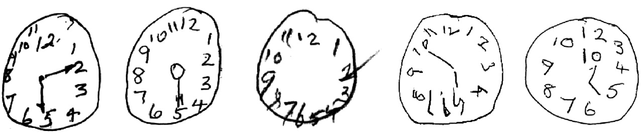
Although deficits in dressing and constructional ability are termed apraxias, they are best considered as visuospatial, rather than motor impairments. Copying three dimensional shapes such as a wire cube, interlocking pentagons, or constructing a clock-face with numbers (fig 2) are good tests of constructional ability, and may also highlight neglect if it is present. Left sided lesions tend to cause over-simplification in copying, whereas right sided lesions may result in abnormal spatial relationships between the constituent parts of the figure. Dressing apraxia is easily tested by having the patient put on clothing that has been turned inside out.

Impaired clock face drawings in dementia.
Visual agnosias
Visual object agnosias cause a failure of object recognition despite adequate perception. Those with apperceptive visual agnosia have normal basic visual functions, but fail on more complex tasks involving object identification and naming. However, they are able to name objects to description, or by touch, indicating a preserved underlying semantic representation of the object. This phenomenon is described with widespread, bilateral occipitotemporal infarction. In cases of associative visual agnosia, the deficit reflects a disruption of stored semantic knowledge, and involves all modalities accessing this information. Lesions of the anterior left temporal lobe are typical. To test for these syndromes, it is necessary to assess object naming and description, along with tactile naming, naming unseen objects to description, and the ability to provide semantic information about unnamed items.
Prosopagnosia
Prosopagnosics cannot recognise familiar faces. Often other clues, such as gait, voice or distinctive clothing, are used to aid identification. The deficit may not be entirely selective to faces, and often fine grained identification within categories may also be impaired (for example, makes of car, types of flowers). Patients are generally able to characterise individual facial features, and since the underlying (semantic) knowledge associated with a particular person is not disrupted, the ability to produce attributes of the face in question, if it is named, remains intact. An inferior occipitotemporal lesion underlies this disability, and is often associated with a field defect, achromatopsia or pure alexia. In delusional misidentification syndromes such as the Capgras syndrome, patients are convinced that an impostor, who looks identical, has replaced a close relative. It occurs in dementia and schizophrenia, and there is a suggestion that the linkage of affective attributes to a face may be disconnected from processing of its identification.
Colour deficits
Colour processing deficits such as achromatopsia (loss of ability to discriminate colours) are often associated with pure alexia after medial occipitotemporal damage, following left posterior cerebral artery infarction. Colour agnosia impairs tasks requiring retrieval of colour information (for example “What colour is a banana?”), and colour anomia (for example “What colour is this?”) refers to a specific disorder of colour naming despite intact perception and colour knowledge, probably caused by a disconnection of the language structures in the temporal lobe from the visual cortex.
A few rare syndromes are worthy of mention. Balint’s syndrome consists of a triad of simultanagnosia (inability to attend to more than one item of a complex scene at a time), optic ataxia (inability to guide reaching or pointing despite adequate vision), and occulomotor apraxia (inability to voluntarily direct saccades to a visual target). Fields may be full when challenged with gross stimuli, and occulocephalic reflexes are intact. This syndrome results from bilateral damage including the superior-parieto-occipital region, which disrupts the dorsal (“where”) visual processing stream linking visual with parietal association areas. Possible causes include carbon monoxide poisoning, watershed infarction, leucodystrophy, and the posterior cortical variant of Alzheimer’s disease. Anton’s syndrome is a visual agnosia, in which the patient denies any deficit and may attempt to negotiate the environment, invariably without success. In the curious phenomenon known as blindsight, visual stimuli can induce a response despite cortical blindness. It is probably mediated by perceptual processing in subcortical structures and brainstem nuclei.
GENERAL NEUROLOGICAL EXAMINATION
A cognitive assessment is incomplete without a careful general neurological examination. There are a number of clinical features that have particular importance. Although in the early stages of many neurodegenerative diseases, clinical signs may be absent, this is not invariable. Table 2 highlights the important neurological findings associated with various cognitive disorders.
Associated neurological features
COGNITIVE RATING SCALES
Perhaps the most widely used cognitive rating scale is the mini mental state examination (MMSE), which although extremely useful, is weighted significantly towards aspects of memory and attention. The language tasks, however, are relatively insensitive, and there is little assessment of visuospatial ability, and no testing of executive performance. It is scored out of 30, with a score of 24 or less being regarded as abnormal. The patient’s educational background, age, and first language should be considered. It is, however, fast to administer and well recognised as a screening instrument.
The Addenbrooke’s cognitive examination (ACE) has been developed in an attempt to address the deficiencies of the MMSE. It was designed to be sensitive to the early stages of frontotemporal dementia and Alzheimer’s disease. The 100 point scale incorporates the items of the MMSE, but includes more tests of executive function, visuospatial skill, and more complex language assessment.
The briefest of all of the screening examinations is the mental test score (MTS), which is a 10 item scale assessing orientation, memory (anterograde and retrograde), and attention. A score of 6 or less is abnormal in the elderly, but as with the other cognitive rating scales, the profile of deficits is more instructive than the global score. It may help direct further, more detailed assessment, but offers no advantages over the other scales other than speed of administration.
FORMAL NEUROPSYCHOLOGICAL ASSESSMENT
Unfortunately, neuropsychological services are not always available. In general it is reasonable to reserve this facility for patients in whom a more detailed assessment is needed for diagnostic purposes, or those in whom one wishes to better characterise deficits for the purposes of rehabilitation. Patients with clear-cut deficits of moderate severity are unlikely to need formal testing to reach a diagnosis. In contrast, neuropsychology has much to offer for patients in whom deficits are early and subtle, or who have marginal performance on cognitive screening scales.
A list of common neuropsychological tests and what they assess is included in table 3.
Selected neuropsychological tests
CONCLUSION
It is impractical to examine everything in the cognitive assessment, and as in most other areas of neurology, the history remains pre-eminent in guiding subsequent examination. The central role of an informant, and the ability to immediately test hypotheses generated during the history taking, distinguish this means of neurological assessment.
In some patients it is not possible to reach a firm diagnosis after a single cognitive assessment, even when armed with a formal neuropsychological report. This is particularly true for the mild stages of neurodegenerative diseases, and reflects the relative insensitivity of both clinical and imaging assessment to early pathology. The time honoured method of longitudinal follow up and repeated assessment in such cases is invaluable, and should not be forgotten.
REFERENCES Cognitive assessment overview
Cognitive domains
Neurodegenerative diseases
Footnotes
-
Dr Kipps is supported by the Wellcome Trust, and Professor Hodges by a Medical Research Conncil Programme Grant.