Article Text

Statistics from Altmetric.com
- neurological intensive care
- stroke
- subarachnoid haemorrhage
- status epilepticus
- meningitis
- Guillain-Barré syndrome
The majority of neurologists work in district general or teaching hospitals with large general intensive care units (ICUs). In this setting, ICUs require an increasing input from neurologists, especially with regard to the assessment of hypoxic brain damage and the neurological complications of organ failure, critical illness, and sepsis. In contrast, dedicated neurological intensive care units (NICUs) tend to deal largely with a different population of patients. Such units are primarily concerned with the management of primary encephalopathic patients, the control of raised intracranial pressure (ICP), the management of ventilatory, autonomic, and bulbar insufficiency, and the consequences of profound neuromuscular weakness. This role encompasses the treatment of mechanical ventilatory failure, specific treatments (both medical and surgical) and general medical complications of these disorders.1
In general, NICU patients with primary neurological diseases such as myasthenia gravis, Guillain-Barré syndrome, central nervous system infections, status epilepticus, and stroke have a better outcome than those patients with secondary neurological disease seen on general ICUs. However, such patients remain dependent on ICU support for very much longer periods of time. This results in very significant psychological demands on the patients, their carers, the nurses, physicians, and other health care professionals. In this review we will consider the rationale for managing acute neurological conditions in a dedicated NICU environment.
INDICATIONS FOR ADMISSION
Indications for admission to NICU include:
-
impaired level of consciousness
-
impaired airway protection
-
progressive respiratory impairment or the need for mechanical ventilation (box 1)
-
seizures
-
clinical or computed tomographic (CT) evidence of raised ICP caused by a space occupying lesion, cerebral oedema or haemorrhagic conversion of a cerebral infarct
-
general medical complications (for example, hyper/hypotension, aspiration pneumonia, sepsis, cardiac arrhythmias, pulmonary emboli)
-
monitoring (for example, level of consciousness, respiratory function, ICP, continuous electroencephalography (EEG))
-
specific treatments (for example, neurosurgical intervention, intravenous or arterial thrombolysis).
Box 1: Neurological indications for mechanical ventilation
-
Failure of central regulation of respiration (apnoea, ataxic or cluster breathing)
-
Inability to protect airway
-
Brain swelling with depressed level of consciousness (Glasgow coma score < 9)
-
Impending neuromuscular respiratory failure (forced vital capacity < 20 ml/kg, tachypnoea, dyspnoea at rest, use of accessory muscles, staccato speech)
Respiratory failure must be anticipated before the emergence of hypoxia and/or hypercapnia. Thus the threshold for intubation is lower in the context of rapidly progressive neuromuscular weakness.
Before examining these groups in more detail it is essential to emphasise that there are general principles of intensive care management common to all units, whatever their specialisation. These include meticulous nursing and medical care and, crucially for our patients, physiotherapy. Early and aggressive physiotherapy intervention (including frequent alterations of limb positioning, passive limb movements, and appropriate splinting) helps to maintain joint mobility and prevents limb contractures and pain while awaiting neurological improvement.
Other aspects of general ICU care include the management of agitation and pain, maintenance of an adequate airway and ventilation, cardiovascular stability, nutrition, anticoagulation, thrombolysis, and raised ICP; these will be discussed in related articles.
Many patients with impaired consciousness or severe neuromuscular weakness are not able to communicate adequately. It is essential that satisfactory means of communication are established as soon as possible. Furthermore, when communication is difficult, the family often represents the patient’s interests and they must therefore have access to medical and nursing staff throughout so that they understand the immediate clinical situation, management, and outlook.
The central and peripheral causes in ventilatory insufficiency or failure which may require admission to the NICU are listed in tables 1 and 2.
Central causes of ventilatory insufficiency or failure which may require admission to the NICU
Peripheral causes of ventilatory insufficiency or failure which may require admission to NICU
Stroke
In contrast to other European countries, admission to an NICU following stroke is relatively uncommon in the UK. However, patients with acute stroke, whether haemorrhagic, ischaemic or venous, require resuscitation and close monitoring in an attempt to prevent the secondary cerebral insults and the subsequent clinical deterioration that results from major systemic derangements. This is best achieved in an intensive care or high dependency environment where physiological monitoring can be efficiently undertaken. Obviously not all stroke sufferers can be admitted to a specialist ICU and the level of care will depend on the availability of local stroke units and the condition and prognosis of the patient.
The principles of assessment and resuscitation from acute stroke are similar regardless of the underlying cause and include:
-
airway management—tracheal intubation is indicated when there is:
-
– impaired level of consciousness (for example, Glasgow coma score < 9)
-
– progressive respiratory impairment or respiratory failure
-
– impaired cough and airway clearance
-
– pulmonary oedema/aspiration
-
– seizure activity
-
– intubation may also be required before diagnostic or therapeutic procedures such as magnetic resonance imaging (MRI) or thrombolysis
-
-
maintenance of adequate arterial blood pressure/cerebral perfusion pressure
-
intravenous fluid management
-
temperature control
-
control of seizures
-
institution of enteral nutrition
-
ICP management
-
medical treatment of complications (for example, sepsis)
-
other management related to the underlying cause (for example, anticoagulation, thrombolysis, evacuation of haematoma, clipping and coiling of intracerebral aneurysms).
Middle cerebral artery occlusion
There is much debate surrounding aggressive therapeutic intervention in patients with extensive infarction caused by acute middle cerebral artery occlusion (fig 1) in whom expectation for functional recovery is low.2,3 Following resuscitation and stabilisation there is evidence for early and aggressive intervention with thrombolysis. Numerous exclusion criteria (including > 3 hours elapsed from stroke onset and widespread early infarct changes on CT scan) currently mean that alteplase is probably only appropriate for a small percentage of patients reaching the NICU. Clinical deterioration following middle cerebral artery infarction is common and associated with cerebral oedema, usually developing between 2–7 days. Oedema and infarction causes a mass effect leading to horizontal and vertical distortion and shift of the brainstem. This change in dynamics may not always being reflected by ICP measurements. The management of this brain swelling is often problematic (see Dunn in issue 74). Osmotic diuretics and hyperventilation are rarely effective and some advocate decompressive craniectomy if extensive cerebral swelling occurs, especially following non-dominant infarcts.5 Other causes of deterioration include haemorrhagic transformation of the infarct, which may produce diencephalic brain herniation, the development of seizures, and systemic factors including congestive cardiac failure, pulmonary oedema, cardiac arrhythmias or pulmonary emboli.

Axial T1 weighted MRI scans showing evolution of middle cerebral artery occlusion causing extensive infarction with mass effect. The appearances after decompressive craniotomy are shown in the third panel.
Acute basilar occlusion
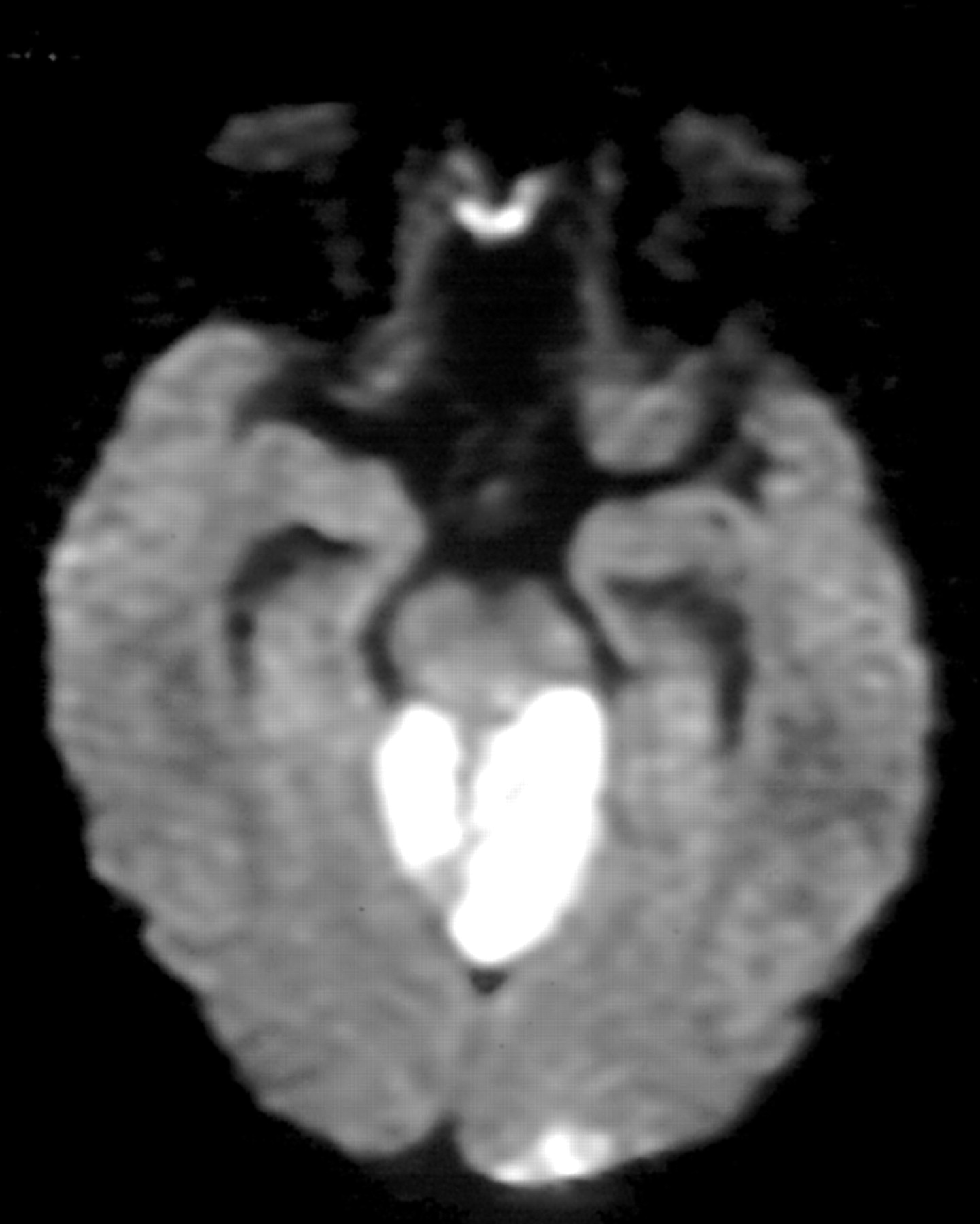
Acute basilar occlusion (fig 2) is a potentially fatal event requiring early recognition and urgent intervention. Prognosis is particularly poor if there is rapid progression to coma with the need for tracheal intubation and ventilatory support. Because of the devastating consequences of established basilar occlusion, aggressive intervention may be indicated in the early stages. Anticoagulation with intravenous heparin remains the mainstay of treatment, but thrombolysis up to 12 hours after onset or continuing progression may be indicated despite the risks of fatal intracranial haemorrhage (although randomised trial data are lacking). Late deterioration occurs in up to a third either because of extension of the thrombus causing successive occlusion of the perforating arteries or due to “artery-to-artery” emboli arising distally from the site of the occluded vessel. The prognosis is usually poor in patients who present with established infarction, but those with limited infarcts of the pons may have a reasonable functional outcome, especially with early thrombolysis or anticoagulation, particularly if admitted and stabilised before the brainstem dysfunction is fully established or mechanical ventilation required.

Axial diffusion weighted MRI scan showing extensive brainstem infarction in a patient with basilar occlusion associated with antiphospholipid syndrome and dehydration.
Cerebellar infarcts
Cerebellar infarcts may be difficult to recognise on account of the slow evolution of brainstem and cerebellar signs. Late deterioration is associated with an increase in infarct volume leading to brainstem involvement and herniation, compression of the brainstem caused by oedema, and the development of hydrocephalus resulting from obstruction of cerebrospinal fluid flow.
Subarachnoid haemorrhage
Admission to NICU is indicated, where possible, for all patients with subarachnoid haemorrhage, to manage systemic complications, recognise and treat clinical deterioration, investigate the cause of the haemorrhage, and to treat any underlying aneurysm or arteriovenous malformation. Resuscitation is directed towards maintaining cerebral perfusion pressure by providing adequate arterial blood pressure (often with the use of inotropes to produce relative hypertension), ensuring a relatively high circulating blood volume (hypervolaemia), and producing relative haemodilution (“triple H therapy”). Other aspects of management in the acute stages include suitable analgesia, seizure control, and treatment with nimodipine to prevent secondary ischaemia caused by vasospasm. Angiography and definitive treatment should be undertaken as soon as possible. Although the relative role and timing of operative clipping and endovascular treatments are still uncertain, it is clear that these patients should be monitored in an intensive care environment. Sudden death may occur in up to 10% of cases because of early rebleeding, intraventricular extension of the haemorrhage, or general medical complications including pulmonary aspiration, cardiac arrhythmias, and neurogenic pulmonary oedema. Clinical deterioration may also develop because of delayed cerebral ischaemia caused by progressive vasospasm, enlargement of intracerebral haematoma or the development of hydrocephalus.6
Supratentorial intracerebral haemorrhage (ganglionic or lobular)
Admission to the NICU will be determined by the clinical state of the patient and the prognosis. Following adequate resuscitation there remains considerable uncertainty about the role of surgical intervention (for example, evacuation of haematomata, drainage of hydrocephalus, decompressive craniectomy), but aggressive treatment may be indicated when monitoring reveals raised ICP unresponsive to medical treatment. Acute deterioration may occur in 30–60% of patients and usually within the first two days; close monitoring is therefore essential. The causes of deterioration include increase in haematoma volume, development of penumbral oedema, obstructive hydrocephalus, or systemic complications such as aspiration pneumonia, sepsis or cardiac arrhythmias.
Infratentorial intracerebral haemorrhage (cerebellar or brainstem)
The acute management will usually involve early tracheal intubation and mechanical ventilation and urgent control of ICP. Clinical deterioration is common and is usually secondary to direct brainstem compression accompanied by cerebellar herniation rather than obstructive hydrocephalus. Rebleeding is a neurosurgical emergency and the high mortality often justifies surgical evacuation.
Cerebral venous thrombosis
Cerebral venous thrombosis is particularly important to recognise. Although robust randomised trial data are lacking, there is general consensus that early anticoagulation can result in good clinical outcome, even in the face of haemorrhagic venous infarction. MR and CT vascular imaging has made it easier to establish the diagnosis, but close monitoring of the patient is essential as late deterioration can have many causes. These include extension of the thrombosis, development of haemorrhagic infarction, raised ICP secondary to cerebral oedema, seizures, the development of systemic complications including aspiration pneumonia, pulmonary emboli, and sepsis, and complications of an often associated hypercoagulable state.
Status epilepticus
Patients with severe epilepsy and status epilepticus are often admitted to general intensive care units. Commonly identified causes of status are medication change, encephalitis, trauma, cerebrovascular disease, tumours or acute metabolic or toxic disturbances. However, NICUs will often have to treat patients with known epilepsy who have developed refractory status, which has not responded to conventional management either because of severe underlying epilepsy or serious cerebral irritation caused by the underlying cause.
Following adequate resuscitation the treatment of status epilepticus in the NICU proceeds simultaneously on four fronts: termination of seizures, prevention of seizure recurrence once status is controlled, management of the precipitating causes, and management of the complications. The drug treatment of status epilepticus has been reviewed recently7–9 and admission to NICU should be undertaken as soon as possible when it is clear either that conventional treatment (that is, lorazepam, phenytoin, phenobarbitone) has failed to abort seizure activity, or if there is significant sedation, failure to protect the airway or ventilatory impairment, or when general anaesthesia is indicated. In addition to monitoring respiratory and cardiac function, continuous EEG monitoring is necessary in prolonged and refractory status. The appropriate titration of anaesthetic agents during status epilepticus may be based on the appearance of burst suppression on the EEG. Furthermore continuous recording will give an indication of worsening of generalised convulsive status epilepticus regardless of the presence or absence of sedating drugs or paralysing agents. It is striking that in a relatively recent survey less than a third of units monitored status by continuous EEG or cerebral function monitor, and almost a half used clinical monitoring only.10
The complications of status epilepticus relate either to the cerebral and metabolic consequences of prolonged seizures or the effects of medical treatment. Cardiopulmonary problems include the development of aspiration pneumonia, adult respiratory distress syndrome, pulmonary emboli, myocardial ischaemia, and cardiac arrhythmia. Hyperthermia is common and rhabdomyolysis may develop. Prolonged hypoxia may cause cerebral damage and electrolyte disturbance, and metabolic acidosis may contribute to the development of multi-organ failure. Many drug treatments used in status epilepticus cause sedation, respiratory depression, and hypotension. Artificial ventilation is required if general anaesthesia is indicated or if the seizures remain difficult to control. It is also necessary to maintain systemic blood pressure at normal or supranormal levels to ensure adequate cerebral perfusion. Fluid resuscitation and/or inotropic support should be guided by appropriate cardiovascular monitoring.
Acute bacterial meningitis
Bacterial meningitis remains a potentially devastating neurological disorder. Although survival rates have improved, the mortality rate of acute meningitis remains significant and there is a high incidence of residual severe neurological deficit, particularly if there is a delay in initiating treatment and monitoring for complications.11 Adult meningitis is commonly caused by Streptococcus pneumoniae (pneumococcus) or Neisseria meningitides (meningococcus). Escherichia coli and Staphylococcus species account for a small number of cases while Haemophilus influenzae occurs in children. Less commonly meningitis may be caused by Listeria monocytogenes and Pseudomonas species, especially in the immunocompromised patient. Tuberculous meningitis may present at any age, and requires a high index of suspicion. Patients with meningitis are usually admitted to an ICU when in coma, when there are complications such as seizures and cerebral oedema, or because they have developed systemic problems including septicaemia, pulmonary aspiration or cardiopulmonary compromise. The ICU environment allows close supervision of supportive and specific treatment.
It is essential to exclude cerebral abscess and subdural or extradural empyema as well as other causes of meningism with appropriate imaging before lumbar puncture is undertaken. If imaging facilities are unavailable, urgent empirical treatment with ceftriaxone or cefotaxime is indicated. The early complications of acute meningitis include the development of cerebral oedema, transtentorial herniation, and coma. Immediate transfer to an ICU is necessary under these circumstances. Bacterial meningitis caused by any organism may result in septic or hypovolaemic shock. Thus, haemodynamic and respiratory monitoring are mandatory at an early stage and mechanical ventilation may be required if there is evidence of cardiorespiratory compromise. Bacterial meningitis is a notifiable disease. Chemoprophylaxis with rifampicin or ciprofloxacin is indicated for all household and healthcare contacts of patients with meningococcal infection.
In adults with normal renal function the recommended empirical treatment for bacterial meningitis consists of intravenous (iv) ceftriaxone 4 g followed 24 hours later by 2 g iv daily, cefotaxime 2 g iv every four hours, benzylpenicillin 1.2 g iv every four hours, or ampicillin 2 g iv every four hours, all of which cover the common pathogens and most enteric Gram negative organisms. If resistant pneumococcus or listeria is suspected or there is a concern about an immunosuppressed patient, vancomycin 2 g iv every 12 hours should be added. If patients have a penicillin allergy chloramphenicol 2 g every six hours is recommended.
Where tuberculous meningitis is suspected treatment should be instituted, and may need to be continued without positive bacteriological evidence for several weeks until the results of cultures are negative. Standard antituberculous treatment consists of isoniazid (20 mg/kg once daily), rifampicin (20 mg/kg) and pyrazinamide (40 mg/kg), together with pyridoxine (10 mg). Second line drugs (streptomycin, ethionamide, and ethambutol) may be required to treat resistant organisms.
The role of steroids in the treatment of adult bacterial meningitis continues to be debated. A recent trial lent support to the use of dexamethasone (10 mg every six hours, started with the first dose of antibiotics) for pneumococcal meningitis. Weaker evidence supports its adjuvant use in tuberculous meningitis.
Late deterioration following acute bacterial meningitis may be caused by antibiotic resistance or to the development of cerebral oedema, subdural effusion or empyema, superior sagittal sinus thrombosis, hydrocephalus, the development of focal neurological signs caused by an associated vasculitis, or systemic complications including pericardial effusion and polyarteritis.
Acute viral encephalitis
Acute infectious encephalitis and parainfectious inflammatory encephalopathy are rare complications of common viral infections. A causal agent is found in less than 50% of cases of viral encephalitis.12,13 The most common cause is herpes simplex virus but other human herpesviruses (for example, varicella zoster), enterovirus, mumps, measles, and viruses associated with respiratory tract infections (adenovirus and influenza B) are also important causes. Encephalitis caused by HIV or related to immunosuppression in AIDS is becoming increasingly frequent.
Viral encephalitis may present with the rapid development of encephalopathy and coma; this often requires tracheal intubation for airway protection and ventilatory support, control of raised ICP, and the effective treatment of seizures. Patients are often confused, restless or aggressive, even in the absence of focal neurological signs, and sedation may be necessary.
The use of polymerase chain reaction for the diagnosis of herpes simplex encephalitis14 and other causes of viral encephalitis (for example, cytomegalovirus (CMV), Epstein-Barr virus, enterovirus) has meant that brain biopsy is usually no longer necessary to reach a definitive diagnosis. The management of acute viral encephalitis includes appropriate airway management, fluid and nutritional support, and treatment of confusion and seizures. Specific treatment for herpes simplex encephalitis with aciclovir (10 mg/kg iv every eight hours for at least 10 days) reduces mortality by 25%, particularly in the most severely affected, with up to 40% of such patients making a good or complete recovery.15 For CMV encephalitis ganciclovir is indicated (10 mg/kg every 12 hours).
Clinical deterioration in herpes simplex encephalitis is usually the result of severe cerebral oedema with diencephalic herniation or systemic complications, including generalised sepsis and aspiration (fig 3). Furthermore, progressive worsening of focal seizures may lead to status epilepticus. The use of ICP monitoring in acute encephalitis remains controversial but should be considered if there is a rapid deterioration in the level of consciousness, and imaging suggests raised ICP. In this situation, aggressive treatment including tracheal intubation and mechanical ventilation with appropriate sedation should be instituted, and seizures treated. Prolonged sedation or general anaesthesia may be necessary. Decompressive craniotomy may be successful in cases where there is rapid swelling of a non-dominant temporal lobe as poor outcome is likely without.

Coronal T1 weighted MRI showing extensive grey and white matter changes with a temporal lobe emphasis in a patient with herpes simplex encephalitis.
Acute parainfectious inflammatory encephalopathy
Acute disseminated encephalomyelitis (ADEM) and acute haemorrhagic leucoencephalitis (AHL) are inflammatory disorders presumed to be the product of an autoimmune response to infection, although a clear history of preceding illness is often absent.16 In AHL and the more aggressive presentation of ADEM, patients may develop rapid and profound deterioration in the level of consciousness and present in coma or with established focal neurological deficits, causing ventilatory impairment or failure to protect the airway.
Although high doses of corticosteroids are commonly given there is no randomised controlled trial evidence for their efficacy, and mortality from AHL is high even if patients are promptly admitted to the NICU. The prognosis for ADEM is rather better and a proportion of patients may make a good recovery, although many are left with significant residual cognitive or focal deficits.
Multiple sclerosis
Patients with multiple sclerosis may develop respiratory insufficiency.17 This is multifactorial and may be associated with respiratory muscle weakness, bulbar dysfunction, and disorders of the regulation of breathing. Acute multiple sclerosis relapses caused by plaques in the brainstem or high cervical cord may lead to acute respiratory and bulbar failure. Admission to the NICU and temporary airway protection and respiratory support, either continuous or confined to the period during sleep, may be necessary during acute bulbar or spinal relapses.
Autonomic neuropathy/movement disorders
Patients with autonomic neuropathy, especially those associated with parkinsonian syndromes (for example, multisystem atrophy) may develop respiratory insufficiency related to impairment of the control of breathing and vocal cord paresis. Admission to the NICU for ventilatory support, either intermittent or continuous, is occasionally necessary. Movement disorders, in particular status dystonicus, may require admission for respiratory or bulbar compromise or physical exhaustion.18
Cervical spinal cord lesions
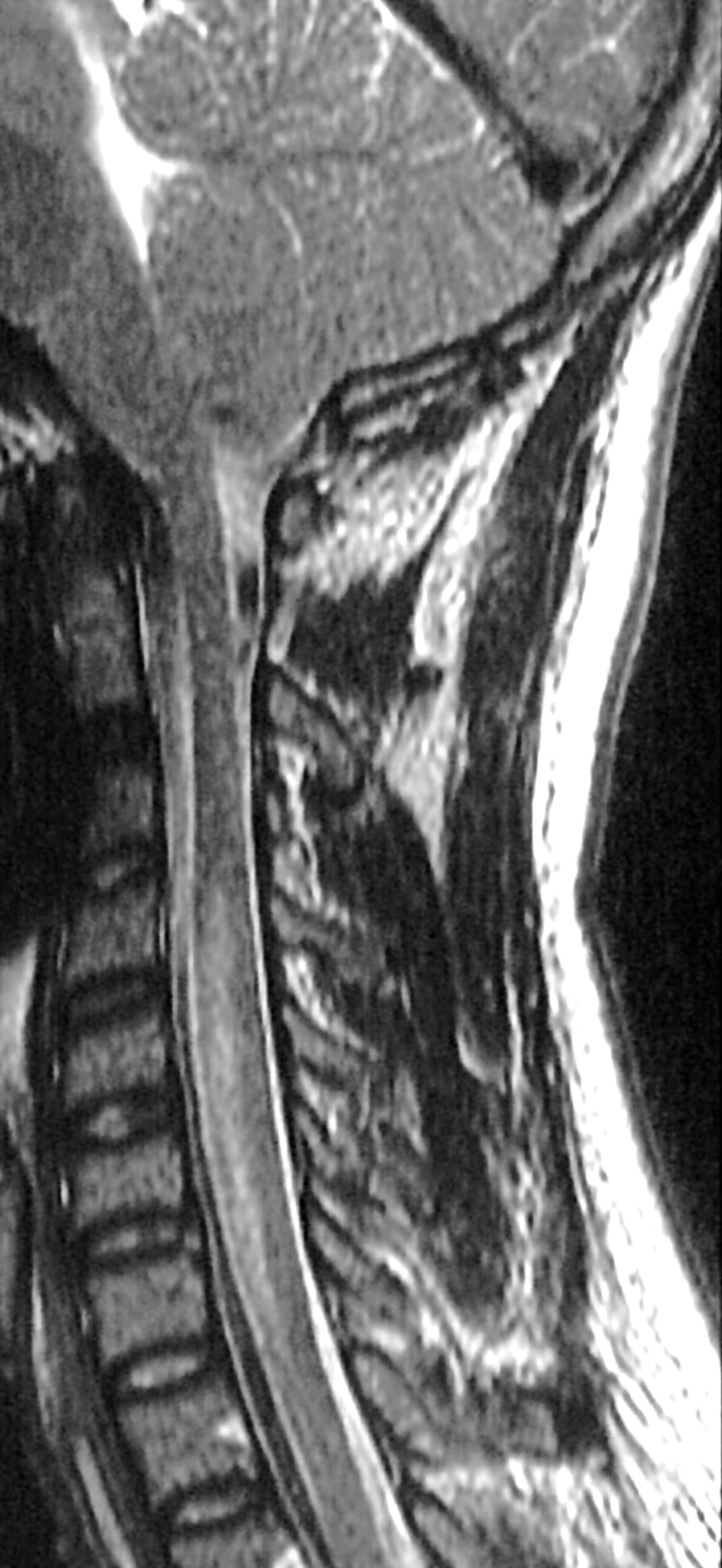
Lesions of the cervical spinal cord may lead to acute respiratory failure requiring ventilatory support; these conditions include postinflammatory myelitis, structural lesions caused by rheumatoid disease, and other skeletal abnormalities at the foramen magnum. Acute deterioration is often precipitated by trauma or intercurrent events (fig 4).
Sagittal T2 weighted MRI scan showing cord infarction causing respiratory failure caused by anterior spinal artery occlusion.
Neuromuscular disease
Patients with neuromuscular disease will require care in an NICU if they develop acute or acute on chronic respiratory failure or acute bulbar (or limb) weakness. The underlying neuromuscular disease may be a primary presentation (for example, Guillain-Barré syndrome) but may also have been relatively stable (for example, post-poliomyelitis), slowly progressive (for example, acid maltase deficiency), more rapidly progressive (for example, motor neurone disease or Duchenne muscular dystrophy) or relapsing or intermittent (for example, myasthenia gravis). Patients with these disorders may develop severe and unexpected deterioration of respiratory, bulbar, and limb function as a consequence of intercurrent events including systemic infection, disease exacerbation, and increased pulmonary load—for example, pregnancy or obesity. It must also be emphasised that patients may develop neuropathy, myopathy or disorders of neuromuscular transmission as a consequence of multiorgan failure, sepsis or prolonged ICU care. These conditions are considered elsewhere in this issue.
The presence of coexisting bulbar weakness may result in a failure to clear retained secretions leading to pulmonary aspiration and the development of bronchopneumonia. Thus patients with neuromuscular disease must be closely monitored for the development of a progressive decline in forced vital capacity, arterial oxygen saturation, and arterial blood gas tensions. Regular chest x rays are often indicated.
Acute poliomyelitis/motor neurone disease/mitochondrial disease
During acute poliomyelitis respiratory insufficiency occurs as a result of respiratory muscle weakness or involvement of the central respiratory control mechanisms. Respiratory insufficiency may develop many years after poliomyelitis, even in the absence of any obvious respiratory involvement during the acute illness or convalescent phase.
Respiratory insufficiency is the common terminal event in motor neurone disease either due to respiratory muscle or bulbar weakness leading to alveolar hypoventilation, pulmonary aspiration, bronchopneumonia or pulmonary emboli. However, some patients with motor neurone disease may develop respiratory insufficiency early in the course of the disease and may even present with respiratory failure or respiratory arrest. Such patients may undergo tracheal intubation and mechanical ventilation, and be admitted to general ICUs before the diagnosis has been made. The progressive nature of the condition means that weaning to non-invasive ventilatory support may be impossible and requires the specialist support of a NICU.
Mitochondrial disease may present to the NICU as status epilepticus, with recurrent stroke-like events, as a metabolic coma caused by unexplained lactic acidosis, or with respiratory failure caused by central or peripheral impairment of ventilation.19
Guillain-Barré syndrome
The role of the ICU in the management of Guillain-Barré syndrome is well documented. The indications for admission include ventilatory insufficiency, severe bulbar weakness threatening pulmonary aspiration, autonomic instability, or coexisting general medical factors. Often a combination of factors is present. The incidence of respiratory failure requiring mechanical ventilation in Guillain-Barré syndrome is approximately 30%. Ventilatory failure is primarily caused by inspiratory muscle weakness, although weakness of the abdominal and accessory muscles of respiration, retained airway secretions leading to pulmonary aspiration and atelectasis are all contributory factors.20,21 The associated bulbar weakness and autonomic instability reinforce the need for control of the airway and ventilation. Acute motor and sensory axonal neuropathy, the acute axonal form of Guillain-Barré syndrome, usually presents with a rapidly developing paralysis developing over hours and the rapid development of respiratory failure requiring tracheal intubation and ventilation. There may be total paralysis of all voluntary muscles of the body, including the cranial musculature, the ocular muscles, and the pupils. Prolonged paralysis and incomplete recovery are more likely and prolonged ventilatory support may be necessary.
Using Guillain-Barré syndrome as an example of the problems of the long term paralysed and ventilated patient the more common complications include:
-
Respiratory tract infections that may be associated with aspiration caused by oropharyngeal weakness but are also common in the paralysed patient as a consequence of nosocomial infections associated with endotracheal tubes or tracheostomy.
-
Cardiac involvement characterised by arrhythmias caused by autonomic impairment, most often tachycardias but more seriously bradyarryhthmias.
-
As with other acute neurological disorders requiring ventilation, mild hyponatraemia is common and usually associated with the syndrome of inappropriate antidiuretic hormone secretion.
-
Gastrointestinal involvement may include the development of paralytic ileus, which may be severe.
-
Confusion is also particularly frequent in ventilated patients and often unrecognised.
Following recovery most patients report that hallucinations, often distressing, occurred during the period of ventilation. The abnormal mental state is multifactorial, probably relating to metabolic and acid base disturbances, drugs, pain, and sensory deprivation. Pain is particularly common in Guillain-Barré syndrome (more than half of patients during admission) but is also a major problem in other acute neurological disorders.
The neurocritical care unit is also the most appropriate place for the provision of immunotherapy—particularly plasma exchange and possibly intravenous human immunoglobulins—and the reduction in morbidity and mortality associated with these treatments is likely to be partly related to the specialised ICU environment in which the treatments are usually given.
Acute ventilatory failure requiring admission to the NICU, intubation, and ventilation may be caused by phrenic neuropathies which will be discussed elsewhere in this issue.
Myasthenia gravis
In myasthenia gravis admission to the NICU is indicated by the development of incipient ventilatory failure, progressive bulbar weakness leading to failure of airway protection, or severe limb and truncal weakness causing extensive paralysis. Admission should be determined by the rate of progression, the presence of bulbar weakness, and the clinical state of the patient rather than an absolute level of forced vital capacity alone. Respiratory failure often results from a myasthenic crisis (usually precipitated by infection, surgery or inadequate treatment), but may also more rarely be precipitated by a cholinergic crisis. The associated bulbar weakness predisposes to pulmonary aspiration and acute respiratory failure necessitating urgent tracheal intubation and ventilation. Expiratory and inspiratory intercostal and diaphragm weakness is common even when there is only mild peripheral muscle weakness. Patients with recent onset generalised myasthenia gravis started on a high dose, daily corticosteroid regimen are particularly at risk for acute paradoxical deterioration during the first 48–96 hours of treatment. Thymectomy should be coordinated by an NICU with experience of the procedure; postoperative management requires such experience.
A number of primary myopathic disorders may present with acute ventilatory failure, but this usually develops when there has been preceding hyoventilation and these conditions will be discussed elsewhere.
Botulism
Botulism must be distinguished from Guillain-Barré syndrome and myasthenia gravis. Patients with acute bulbar and respiratory impairment require admission to the NICU. There may be blurred vision, diplopia, ptosis, dysarthria, dysphagia, and progressive descending limb weakness. Mechanical ventilation is often required. Prominent gastrointestinal and autonomic symptoms in addition to weakness are indicative of botulism, but definitive diagnosis requires detection of toxin in the patient’s serum, stool or food. Treatment consists primarily of supportive care. Antitoxin may be helpful, particularly in type E botulism, but in most adult patients treatment with antitoxin does not lead to significant improvement. If patients survive the acute phase of illness, recovery is usually complete. Mortality in the era of the ICU is less than 10%.
Tetanus
Most patients with tetanus will be admitted to an NICU because of increased muscle tone and spasms which typically begin in the masseter muscles, resulting in the classic finding of trismus. Respiratory compromise is caused by spasm of respiratory muscles or laryngospasm. Autonomic dysfunction occurs in severe cases and results in heart rate and blood pressure lability, arrhythmias, fever, profuse sweating, peripheral vasoconstriction, and ileus. Muscle rupture and rhabdomyolysis can complicate extreme cases. Treatment of tetanus includes removal of the source of the toxin, through wound cleaning and debridement. Human tetanus immunoglobulin neutralises circulating toxin but has no effect once the toxin is neural bound, and should therefore be administered early. Supportive care consists of treatment in a quiet ICU setting to allow cardiorespiratory monitoring with minimal stimulation. Benzodiazepines are used to control muscle spasm and large doses may be required. Therapeutic paralysis with neuromuscular blocking agents may be necessary in severe cases. Intubation may be required owing to hypoventilation caused by muscular rigidity or laryngospasm and should be performed in a controlled, elective manner if possible. Profound muscle rigidity increases energy needs and insensible fluid losses, and additional hydration and nutritional supplementation are required. Treatment in the ICU has resulted in a pronounced improvement in prognosis for patients with tetanus. Modern mortality is approximately 10%. Severe muscular rigidity may last for weeks, with mechanical ventilation required for up to 3–4 weeks. A complete recovery is typical, although mild painful spasms can persist for months.
THE ROLE OF THE NEUROLOGICAL ICU
The primary role of the NICU is that of the management of acute neurological emergencies. This involves control of airway, respiratory, bulbar, and haemodynamic compromise. It also entails the provision of specific treatments and the prevention and management of the secondary complications. In order to achieve this aim, meticulous monitoring of cardiorespiratory variables is essential, reinforced by the specialist skills of intensive care nursing.
Abbreviations
-
ADEM: acute disseminated encephalomyelitis
-
AHL: acute haemorrhagic leucoencephalitis
-
CMV: cytomegalovirus
-
CT: computed tomography
-
EEG: electroencephalogram
-
ICP: intracranial pressure
-
ICU: intensive care unit
-
MRI: magnetic resonance image
-
NICU: neurological intensive care unit
Apart from facilitating medical monitoring and treatment, what other advantages accrue from the centralisation of patients with neurological critical illness as opposed to management of individual cases in scattered general medical intensive care units? Primarily does it alter prognosis? This is extremely difficult to prove with any certainty because of the relative infrequency of the disorders treated and the difficulty in gathering adequate data from ICUs with disparate patient populations. Certainly specialist units will have greater experience in the anticipation, early recognition, and management of potentially fatal complications. In any case the centralisation of these critically ill patients allows the development of care protocols and potentially provides an important role in auditing practices against such established protocols.
Training is another important issue. In the USA and Europe the techniques for caring for critically ill neurological patients have been integrated into the mainstream of neurological and neurosurgical training programmes. The development of a neurocritical care research consortium across Europe, North America, and Japan may be an extremely important development in coordinating research and trials across different units.
Finally, to consider the most difficult area—economic and ethical aspects—it is clear that the use of intensive care varies widely and it has proved difficult to show that greater availability of intensive care facilities leads to better outcome. The role of the NICU should be to formulate and implement worthwhile guidelines to ensure patients with neurological disease treated in any intensive care are effectively managed. The development of NICUs is likely to permit a more efficient application of these techniques to support and treat patients who are critically ill. The aim is to prevent unnecessary morbidity and mortality while maintaining the pastoral aspects of care.